
Optical Microscopy: Considerable Life in Centuries-Old Method
Scientists have used light microscopes for hundreds of years
In laboratory environments where advances in science and instrumentation are taken for granted, the idea of stepping backward seems counterintuitive. Yet that is what biologist David Nguyen, PhD, argues for in his article, “The Advantages of Studying Cells Under a Light Microscope.” Nguyen begins by flat-out stating that the “compound microscope is the most cost-effective type of microscope, making it the preferred choice for many life science applications.”
From their names alone one would guess that light or “compound” microscopes—of the type that readers first laid eyes on in high school biology class—are simpler, less expensive, and easier to use than atomic force or scanning electron microscopes.
Today’s most advanced imaging platforms provide more information per experiment, in less time, than light microscopes, but their price tags are a hundred- to a thousand-fold higher. It is this value argument on which Nguyen makes his case for light microscopy.
Scientists have used light microscopes for hundreds of years and developed tens of thousands of methods based on them for studying organs, tissues, and individual cells. “The combination of low cost and the large amount of biological information that a light microscope provides makes it an invaluable tool for research and medicine,” Nguyen writes.
Optical stains used to visualize features in cells persist long after fluorophores have faded. Light also provides a sort of sweet spot in terms of macro-to-micro field. For example, a pathologist can study the rough texture of cancerous tissue with a light microscope and then, with a flick of the objective lens and refocusing, easily probe the structure’s individual cells, and with the aid of various stains, home in on organelles or study the cell’s functions or life cycle.
Optical tweaks and modes
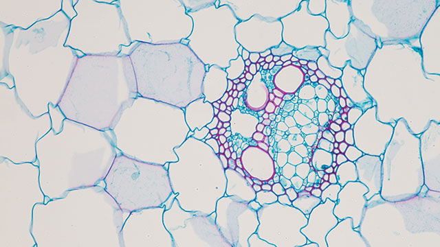
That is not to say that optical microscopy is now and forever in its most advanced state. For example, researchers recently reported on a technique that tweaks optical microscopy in a way that assists in imaging the finer aspects of living plant cells.
Plant cells are among the most studied organisms, but complex plant structures disturb light passing through them. Living plant cells are thick, containing colored and fluorescent details that are often destroyed or altered by the microscope light. That is why light microscopy of live plant cells must balance image quality and information content.
Yosuke Tamada and Masayuki Hattori of Japan’s National Institute for Basic Biology (Okazaki) applied adaptive optics (AO), a technique developed for astronomy to correct for atmospheric disturbance in viewing far away objects. AO improves optical observations by compensating for “wavefront distortions” using a mirror.
AO is most commonly applied to microscopic examination of animal tissues. For plant cell observations, the researchers devised a type of AO that corrects the disturbed light and enables fine imaging of both plant cell organelles and fluorescently-labeled beads attached to leaf cells. Investigators are looking into another technique from astronomy, 3-D AO, to improve even further the ability to visualize processes within plant cells.
AO has also been applied to detailed observation of cellular life cycles. Physics Nobelist Eric Betzig of the Howard Hughes Medical Institute (Loudoun County, VA) combined AO with another old but (until recently) unappreciated technique, lattice light sheet microscopy, to create a “movie” of immune system cells migrating within the ear of an embryonic zebrafish.
Light microscopy will always be constrained by the properties of light and the limitations of optics, but the boundaries of what is possible continue to shift. Tandem methods combining two or more microscopic techniques are one way to continue pushing the limits.
For example, a group at Oxford University led by Robert Ishmukhametov has developed a small, inexpensive addon that confers on optical microscopy four epiillumination methods: back-scattering darkfield (BSDF), epi-fluorescence (EF), interference reflection contrast (IRC), and darkfield surface reflection (DFSR). In contrast to diascopic (transmittedlight) microscopy, epi-illumination is the term for various techniques based on reflected-light microscopy of opaque substances, which include many living cells. Epi-illumination is often used as a fluorescence method with hard materials, but Ishmukhametov found a way to apply it to living cells.
In BSDF mode, the device detected malaria parasites inside human erythrocytes without staining. An EF-based experiment visualized stained Leishmania parasites without the use of excitation filters. IRC and DFSR are used mostly for materials analysis.
Stepping backward
What’s curious about light microscopy is the number of techniques that combine it with more advanced microscopy methods to acquire microscope images containing more information than either instrument alone could produce.
Last year, a group at Switzerland’s Lausanne Polytechnic combined light and atomic force microscopy (AFM) to study the cell cycle in Mycobacterium tuberculosis, the bacterium that causes tuberculosis in humans. In an article published in Nature Microbiology, Dr. Georg Fantner and co-workers reported on the hybrid microscope system, which is clearly the consequence of necessity being the mother of invention.
Rod-shaped bacteria typically divide by splitting around their middles after DNA replication. A mechanism, nucleoid occlusion, protects genes, and a second mechanism, “minicell,” localizes the division site. M. tuberculosis doesn’t use these mechanisms, so scientists interested in studying its cell cycle must resort to novel imaging tactics—in this instance combining light with AFM.
“The AFM provides very high, nanometer-scale resolution, 3-D topography information of the outside of the bacterium, and the ability to measure mechanical properties and perform nanomanipulation,” Fantner tells Lab Manager. “The optical microscope allows you to see inside the cells, and with fluorescently labeled markers provides molecular specificity.”
Being able to look inside the cell with the help of fluorescent tags, he stresses, is the primary advantage of light microscopy for cellular analysis. “We also use this method for mammalian cells, to track their development during proliferation.”
In their paper, Fantner and co-workers noted that the M. tuberculosis study was the longest continuous AFM experiment ever performed on growing cells. Fantner explains that “performing long time experiments with AFM is difficult, especially on living cells. We needed to do long-term experiments to track single bacteri[um] through multiple generations.”
For additional resources on optical microscopy, including useful articles and a list of manufacturers, visit www.labmanager.com/optical-microscopy