

FIB-SEM: A Versatile Nanoscale Laboratory Tool for Advanced Material Science
Achieve a new level of 3D analysis through multi-modal characterization
Scanning electron microscopes (SEMs) are valuable laboratory tools for high-resolution imaging and site-specific surface analysis (topography and composition). Combining an SEM with a focused ion beam (FIB), which provides site-specific milling, deposition, and sputtering of materials, extends imaging and analytical capabilities into the third dimension—depth, and greatly expands the usefulness of both techniques. Other techniques can be added to the FIB-SEM system to provide truly multi-modal characterization, including energy dispersive spectroscopy (EDS), electron backscatter diffraction (EBSD) analysis, Raman spectroscopy, and time-of-flight secondary ion mass spectrometry (ToF-SIMS). These extensions are especially valuable for busy, multi-disciplinary laboratories requiring a wide range of analytical capabilities to support their researchers’ needs.
Combining FIB and SEM brings the power of 3D to the lab
SEM scans the surface of the sample with a finely focused beam of electrons. The beam is generated in an electron column, which gathers electrons from a source, accelerates them toward the sample, and focuses them into a tiny spot at the sample surface. The electrons interact with sample atoms in various ways to generate multiple signals. The SEM synchronously maps point-to-point signal changes into a 2D image as the beam scans a rectangular pattern on the sample surface. Different signals have different characteristics and carry different information. For example, secondary electrons (SE) offer very high spatial resolution and are sensitive to surface topography. Backscattered electrons (BSE) have less spatial resolution but carry compositional information reflecting the average atomic number of sample atoms at each point. Characteristic X-rays have still lower resolution but carry more specific information about elemental composition. The signal, or signals, used depends on the requirements of each analysis.
FIB-SEM adds an ion column to the SEM system, providing a focused ion beam, which can be scanned over the sample in a variety of patterns. FIB can generate images directly, though at lower resolution than SEM. Ions interact with the sample differently than electrons, so FIB images have different contrast mechanisms and can reveal different information.
The primary contribution of FIB, when combined with SEM, is its ability to remove material from the sample. The ions in the beam are much more massive than electrons. On impact, they can eject atoms from the sample—a process called sputtering or FIB milling, allowing the operator nano-scale control to cut precisely positioned cross sections, and reveal sub-surface features. Subsequent SEM analysis of the exposed features delivers high-resolution imaging. Analysis of a sequence of cross sections can generate high-resolution reconstructions of a 3D volume.
FIB-SEM’s ability to shape, polish, or prepare material provides other attractive capabilities, such as milling from both sides that can prepare ultra-thin electron-transparent lamella for analysis in a transmission electron microscope (TEM). Techniques also exist for depositing material with nano-scale control, which is useful for protecting adjacent regions from erosion by the ion beam during delicate milling procedures and also opens the possibility of FIB-generated nano-prototypes.
Combined with other techniques, FIB-SEM can provide the multi-modal analytical capabilities needed in a busy material science lab: EDS for elemental composition, EBSD for crystallographic analysis, Raman for chemical/molecular analysis, and ToF-SIMS for exquisitely sensitive compositional analysis.
Bringing EDS and EBSD microanalysis into 3D
When incident beam electrons displace an inner-shell electron from a sample atom, an electron from a higher energy shell may drop down to fill the vacancy, generating an X-ray with a specific amount of energy that is characteristic of the emitting atom. An EDS spectrometer measures and counts those X-rays, acquiring a spectrum that can determine the quantitative elemental composition of the sample.
EBSD analyzes beam electrons that have been backscattered by the nuclei of sample atoms. The backscattered electrons are diffracted by crystallographic planes in the sample and form a pattern that can be recorded. Deconvolution of the pattern reveals the crystallographic structure of the sample.
An example of FIB-SEM-based EDS and EBSD comes from research at the Ecole Polytechnique in Montreal, where researchers sought to understand the formation of precipitates in additively manufactured high-strength steel and their connections to grain boundaries. The study used serial FIB sectioning, with EDS and EBSD analysis of each section, to generate a 3D representation of the sampled volume. The multi-modal analysis, with EDS revealing the precipitates based on their composition and EBSD showing the grains based on their orientation, allowed the researchers to correlate the two characteristics. The parameters measured included the composition of the precipitates and the size, orientation, and aspect ratio of the grains. The large number of grains and precipitates analyzed provided high confidence in the results.

Adding Raman and ToF-SIMS for multi-modal analysis
Other capabilities for FIB-SEM-based multi-modal material characterization include Raman and ToF-SIMS. Raman spectroscopy looks at the interactions of photons with vibrational modes that characterize intra-molecular bonds. In ToF-SIMS, the secondary ions sputtered from the sample by the FIB are identified by their charge/mass ratio. These techniques were recently used to model degradation mechanisms in lithium-ion batteries (LIB). The investigators developed models for capacity loss based on the cracking and separation of particles that occur during battery cycling. High-resolution imaging from FIB-SEM provided information about morphology and structure. Raman spectroscopy provided information about the state of lithiation, bonding, and disorder in battery materials. ToF-SIMS provided information about the local Li content. Because the FIB can be scanned across the sample, it can measure the Li content at each point to determine its spatial distribution—important information in understanding and validating the degradation model. In this study, the FIB-SEM multi-modal analysis with ToF-SIMS and Raman validated the model relating loss of capacity to degradation of the lithium-ion battery materials.
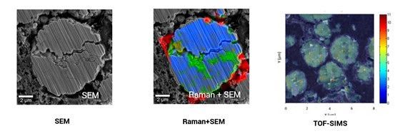
The integration of Raman into the FIB-SEM is accomplished using a high precision stage, which translates the sample from a position under the electron and ion beams to a position beneath the Raman system, which is confocal and scans the sample surface, collecting a spectrum at each point. The high-precision stage permits precise correlation between the structural information obtained by SEM and the spectral information acquired by Raman. The technique is known as Raman imaging and scanning electron (RISE) microscopy. ToF-SIMs integrates easily on a FIB-SEM since the primary ion source needed to produce sputtered secondary ions is already present.
In summary, FIB-SEM provides the opportunity to bring true multi-modal micro and nanoscale 3D characterization techniques into the laboratory for advanced studies in all fields of science or applied research, including materials research, microelectronics, geology, and life sciences.