
Imaging Systems for Biology: Responding to Multidisciplinary Needs
Advances in computer hardware and software, data storage and processing, optics, systems and instrumentation, labeling agents, and reagents have all contributed to the current surge in imaging in the life sciences.
“The widespread use of fluorescent proteins has in particular had a tremendous impact,” says Magnus Persmark, PhD, product manager at Life Technologies (Carlsbad, CA). “We have had leapfrogging in these various areas, for example bioinformatics, reagents, and instrumentation.”
Continuous improvement has been driven by translational and interdisciplinary research that defines modern biology. Rare is the biologist who can make a career from one type of project. Even rarer is remaining within one well-defined area, say, transcription in lower organisms. As biological complexity has revealed itself, imaging tools have kept up. “We now have the technologic foundation to explore connectivities on a cellular and multicellular level in ways that we have never had before,” Persmark adds. “We’re able to visualize complex cell models, spheroids, mixed cell populations, and stem cells, with the ability to label cells and components, their structure and function, and follow that in time and space.”
For imaging, then, necessity has been the mother of invention. New luminescent tags emerge almost weekly that push the limits of detectors and image processing systems, which in turn spawn novel methods and experiments. “At the same time imaging is becoming democratized,” Persmark comments. “With instruments designed for nonexperts entering the market, researchers can more immediately access and assess these new experiments.”
Out with the old
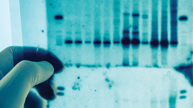
Labs store data from most electrophoresis gels and thin layer chromatography data as standard photo images, providing the media hold their stain or immobilized bands absorb or reflect ultraviolet strongly. A bit more finesse is required for gel bands detected through enzymatic chemiluminescence. “In those situations, you need a camera that will stare at the sample and collect electrons until the enzyme runs its course, which can be 15 minutes or more,” explains James Joubert, application scientist at Photometrics (Tucson, AZ). “The camera should have very low dark current, high quantum efficiency, and extremely low noise so as not to build up a lot of spurious electronic signal.”
Modern biology imaging is nearly synonymous with advanced microscopy, whose macro components are remarkably constant across biology, materials science, and forensics: a light source, a collector/magnifier (usually a microscope), and a collector (camera). Size domains are similar as well, as defects in semiconductors are approximately as large as biology’s top subject matter, cells. Materials and structural imaging differs in its upper size domain, which is sometimes many feet. Within and across those disciplines, however, considerable difference exists in excitation mode and energy. For example, probing into semiconductor material or tissue requires greater power than does skimming surfaces for dopants, or cells for receptors.
Imaging biological systems is arguably the most challenging due to the complexity and dynamism of cells and tissues, and the size and sensitivity dynamic ranges involved. Following single molecules through cells, for example, often requires imaging of surrounding cellular structures that may respond well to white light, while simultaneously measuring rapid fluorescence from deep inside the cell.
“Within biology alone there is such a swath of things, from whole organisms to large tissues, cells, organelles, and molecules,” notes Joubert. His company specializes in charge-coupled device (CCD), electron multiplier CCD (EMCCD), and multichannel imaging systems that all rely on microscopy.
The application list for biology reflects the breadth of applications: digital image restoration, FISH, fluorescence imaging, fluorescent speckle microscopy, FRET, green fluorescent protein imaging, ion imaging, surface trafficking, FRAP, sequential color imaging, single-molecule fluorescence, TIRF—and those are just the microscope-based techniques.
Democratization
For more than a decade, advanced confocal imaging systems provided high-end, high-content visualization of assays and screens. Models from GE Healthcare Life Sciences, PerkinElmer, Molecular Devices, Thermo Scientific, and Olympus cost anywhere from $100,000 to $750,000 —well beyond the budgets of many labs.
Subsequently, GE and Life Technologies pushed cell imaging technology down to a lower price point with, respectively, the Cytell and EVOS instruments that cost around $50,000. Notes Eric Matthews, Midwest sales manager at BMG LABTECH (Cary, NC), “You can think of these as benchtop automated microscopy for cell imaging.”
The next evolutionary phase involved product debuts from BioTek Instruments, Molecular Devices, and PerkinElmer— microplate readers that incorporated automated microscopy functions. “So for a little more than those other entry-level imagers, you could get plate reading and imaging in the same box,” Matthews says. The Cytation from BioTek, SpectraMax from Molecular Devices, and PerkinElmer’s EnSight are actually multimode plate readers with well-imaging capability.
BMG LABTECH does not manufacture such an instrument, but has been “thinking of its pros and cons,” according to Matthews. “They allow you to do pretty advanced imaging, for example, protein co-localization, cell motility, morphology, cell counting— as you can do with any microscope, but in automated format.”
At issue is whether labs should own two standalone instruments or one combination system that is somewhat less expensive. Assuming the combo is the only plate reader in the lab, it’s easy to envision workflow and utilization bottlenecks. “Maybe you have to read two plates and it takes 90 minutes. Meanwhile, your technician can’t read basic protein assays and ELISAs.” In those situations, Matthews says, lab managers may wish they had purchased standalone imagers.
But he’s quick to point out the “obvious advantages” of a dualmode imager/reader. Economics is one plus: Labs may have $80,000 for the combined system but not $110,000 for two instruments. “Plus, you have the ability to do more. Just a few years ago your only option was a very large, half-million-dollar box.”
These systems are also a gateway for companies that previously could not afford imaging except through a core facility. But for these labs, plate readers are already essential workflow components. “Everyone needs a plate reader, so if your lab needs one, and by adding another $20,000 to $30,000 on top of the cost you can get imaging as well, it all seems more doable,” Matthews explains.
As noted, BMG does not have a dual-mode product, but plans to investigate pitching stand-alone readers and imagers with a corporate partner on the imaging side. Matthews views imaging as another wrinkle in the evolution of microplate readers. “Every five to eight years it seems a new mode comes to plate reading, for example fluorescence. Today’s interesting new mode is imaging.”
For additional resources on imaging systems for biology, including useful articles and a list of manufacturers, visit www.labmanager.com/imaging