
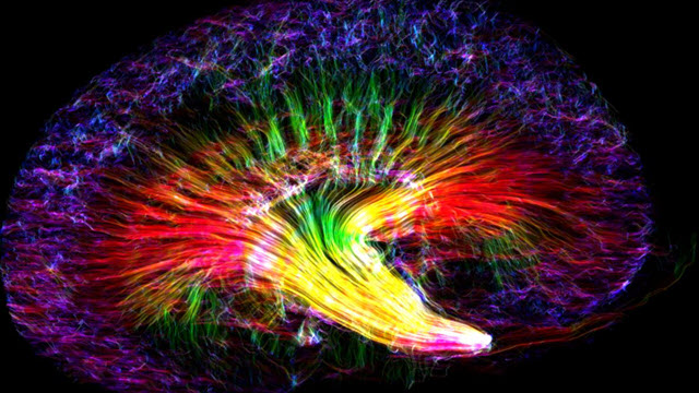
 "Kidney Rainbow"Credit: Nian Wang, Center for In Vivo Microcopy (Directed by Dr. G. Allan Johnson), Duke University, USA via Biomed Central
Earlier in the year, BioMed Central (BMC) launched the second annual BMC ‘Research in progress’ competition, following the success of the inaugural competition in 2017.
The winning image of a mouse kidney has been obtained by diffusion tensor imaging, an MRI-based imaging technique. The bright neon colors represent the orientation of different tubules, which collect filtrate from blood passing through the kidney and process it into urine.
Speaking to BMC about his winning entry, Nian said: “It’s my great honor to receive this award. The image shows the complex 3-D tubular structures of a mouse kidney. It was taken at the Center for In Vivo Microscopy (Directed by Prof. G. Allan Johnson), where our research focuses on developing novel MRI methods to detect tissue microstructures. The non-destructive nature of MRI and its ability to assess the renal microstructure in 3-D make it a promising tool to understand the complex structures of the renal system.”
To read the full article, the runner up, and a selection of entries that caught the eyes of the BMC judging panel, click here.


