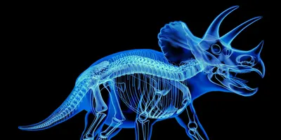
X-ray technology is a foundational tool in both medicine and paleontology, but recent innovations are revealing a surprising intersection between these two fields. Structural biologist Joseph Orgel and his research team at the Illinois Institute of Technology have developed a groundbreaking micron-scale X-ray imaging technique that bridges the past and present—originally created to study fossilized dinosaur tissues, it's now being applied to understand traumatic brain injuries (TBIs) at the cellular level.
TBIs result from sudden trauma to the head, common in sports, vehicle accidents, and military combat. These injuries can cause permanent damage or even death, but they remain difficult to study due to the inaccessibility of deeply embedded neural structures. Orgel's research, presented at the 75th Annual Meeting of the American Crystallographic Association, illuminates a new frontier in biomedical imaging by drawing direct parallels between fossil preservation and neural protection mechanisms.
Preserved Collagen in Dinosaur Fossils Reveals Ancient Structural Insights
The Collagen Conundrum
Orgel’s initial inquiry focused on a paleontological mystery: How have some dinosaur fossils retained collagen, a protein fundamental to connective tissues, for tens of millions of years? Collagen is typically prone to degradation, yet certain samples from the Cretaceous period, including a 68-million-year-old Tyrannosaurus rex, contained remarkably preserved fragments.
The difficulty lay in accessing the protein without destroying the fossil. Traditional methods lacked the sensitivity to differentiate collagen from the surrounding mineralized matrix. This limitation inspired the development of a more sensitive X-ray beam scanning technique capable of resolving structures at the micron scale.
“We had to push ourselves to better develop our X-ray scanning methods,” said Orgel. “Using micron-sized X-ray beams to scan the fossil fragments, we learned how to home in on faint signals of preserved structure.”
The result was a powerful imaging method that could identify tiny, ordered regions of collagen embedded deep within fossil material. These regions, it turns out, were designed to protect more fragile biological structures—a finding that hinted at deeper biological functions with contemporary relevance.
Advanced X-ray Techniques for Analyzing Dinosaur Fossils and Brain Tissue
Orgel’s technique involves using highly focused micron-scale X-ray beams, a significant enhancement over traditional imaging, which often uses larger beams incapable of isolating such faint signals. The advantage lies in its ability to detect ordered molecular structures, particularly the alpha-helix architecture of collagen, without sample destruction.
This method integrates:
- Micro-focus beamlines at synchrotron facilities
- Crystallographic data processing for pattern recognition
- Spectroscopic validation of proteinaceous material
These tools enabled researchers to distinguish between structurally intact and degraded regions, confirming that certain collagen domains serve as biological shields.
Implication for Fossil Preservation
The collagen was most preserved in regions serving structural or protective roles. These biologically reinforced areas safeguarded internal information, akin to how DNA is protected by histones** in modern cells. The findings suggest that structural protection is a core feature of both evolutionary design and post-mortem preservation.
“We realized that the best-preserved regions were not just tough—they were physically shielding the vital ‘instruction manuals’ for cellular interaction,” Orgel explained.
Using Dinosaur-Inspired X-ray Imaging to Study Brain Trauma
Understanding Brain Trauma with Fossil-Inspired Methods
Seeing the success of their technique with dinosaur fossils, Orgel’s team pivoted to apply it to neuroscience, particularly to study myelin sheaths that insulate nerve fibers in the brain. Myelin plays a critical role in neuron function and is vulnerable to damage during traumatic brain injuries.
Using their advanced X-ray imaging method, researchers could visualize myelin damage in unprecedented detail. This enabled the development of a "dose-response" model for brain injuries, identifying the threshold at which physical force leads to irreversible structural damage.
“We can now detect the precise threshold of force that causes permanent structural damage to the myelin sheath that insulates neurons,” said Orgel.
Practical Applications
- Improved TBI diagnostics: Early-stage damage can now be identified before symptoms become severe.
- Helmet design optimization: Data can inform engineering strategies to reduce the risk of brain injuries in athletes and military personnel.
- Therapeutic monitoring: Tracking healing or degeneration at the cellular level could revolutionize rehabilitation programs.
X-ray Insights Beyond Dinosaurs: Heart Valves, Tendons, and Tissue Integrity
Encouraged by their results, Orgel’s team expanded the scope of their imaging technique to include other biological tissues such as Achilles tendons and heart valves. These tissues, like collagen and myelin, rely on fine-scale structural integrity for function.
X-ray imaging revealed subtle damage patterns invisible to standard methods, offering new avenues for early detection of degenerative conditions such as tendinopathies and valvular heart disease. This reinforces the principle that techniques designed for ancient fossils can have profound utility in modern clinical science.
“Seeing the methods we refined for dinosaur bones reveal the invisible damage in brain, muscle, and heart tissues was a true ‘eureka’ moment,” said Orgel, “highlighting the unpredictable and interconnected nature of science.”
Final Thoughts on How X-ray Imaging from Dinosaur Fossils is Transforming Brain Injury Science
What began as a quest to understand the biochemical resilience of dinosaur collagen has blossomed into a transformative approach for diagnosing and understanding human disease. Orgel’s micron-scale X-ray technique, rooted in paleontology, now holds promise for improving neurological diagnostics, enhancing sports medicine, and informing biomedical engineering.
This journey—from fossilized giants to living brains—underscores the vast potential of cross-disciplinary innovation. For laboratory professionals, the implications are clear: combining advanced imaging, materials science, and biology can yield tools that not only unlock the past, but actively shape the future of human health.


