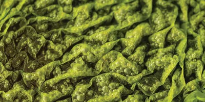
Biofilms are multicellular microbial communities made of bacterial cells surrounded by a network of secreted biopolymers. Biofilms enable bacteria to attach on surfaces and the extracellular matrix barrier structurally isolates bacterial cells from external environmental fluctuations. For instance, biofilms can confer bacterial cell protection against mechanical stress and toxins like antimicrobials and disinfectants.
The word “biofilm” tends to be associated with persistent infection in health care settings such as on dental and bone implants. Biofilms that form on surfaces like water pipes can also adversely affect water quality. However, biofilms are in fact double-edged swords. Beneficial biofilms are being developed as agents to enhance crop protection by acting against phytopathogens, bioremediation treatment of hazardous pollutants, and prevention of corrosion on pipes. This is possible by carefully selecting the types of bacterial communities to exist in a biofilm coating.
Biofilms can be viewed as a community of phenotypically heterogenous cells organizing themselves across different spatial regions and over time to form distinct microenvironments. While the bacterial cells are genetically identical, their different phenotypes within biofilms allow the communities to respond differently to nutrients and oxygen levels, as well as threats. This is an intelligent strategy exploited by both gram-negative and gram-positive bacteria for survival.
“Beneficial biofilms are being developed as agents to enhance crop protection by acting against phytopathogens, bioremediation treatment of hazardous pollutants, and prevention of corrosion on pipes.”
The need for structural characterization of biofilms
There is great interest to capitalize on phenotypic heterogeneity in biofilms to prevent deleterious biofilm formation on implants and to design biofilms as preventive coatings. Currently, spatiotemporal gene expression analyses are the most common methods used to understand biofilm-associated phenotypic heterogeneity at the molecular level. However, it remains unknown how the elemental and structural properties of biofilms differ across space and time because most studies digest biofilms before analyzing the isolated molecules for their quantities.
X-ray diffraction (XRD) is a technique that helps to determine the structures of molecules present in a sample using electron densities of diffracted X-rays. X-ray fluorescence (XRF) is a method used to detect emissions of characteristic fluorescent X-rays from a material that is bombarded with high energy X-rays for elemental analysis.
In a recent study, Azulay et al. made use of XRD and XRF to scan intact B. subtilis biofilm samples of different ages and found spatiotemporal division of four main components including water, spores, extracellular matrix, and metal ions that show how these components interact to affect processes like sporulation and polymer fibril formation.
“Our initial goal was to study the formation of calcium carbonate in whole biofilms, but then we realized that the biofilms themselves, even though they are soft matter organisms, still convey a structural fingerprint. We then adjusted our goal to the study of the dominant structural features in biofilms, the distribution of metal ions, and their relationship with the extracellular matrix properties and biofilm physiology. In this study, we made a major finding that metal ion distribution coincides in space with water channels in biofilms, raising a possible hypothesis on the role of metal ion accumulation in local sporulation along water channels. Additionally, we found that the extracellular protein, TasA, is composed of a weakly repeating cross beta sheet structure. This finding stands in contrast to previous reports of its amyloid nature,” says Liraz Chai, PhD, at the Hebrew University of Jerusalem.
Detecting different ‘types’ of water in biofilms
Using XRD, Azulay and co-workers found that water is bound in biofilm wrinkles, and this demonstrates the role of wrinkles acting as water channels for nutrient transport and waste removal. The team first characterized a mature (10-day-old), naturally hydrated biofilm using XRD, which not only differentiated different polymers and DNA, but also free water and water bound to extracellular matrix and biopolymers. This is supported by a previous study showing that biofilm wrinkles could serve as channels filled with water to transfer nutrients across biofilms, and the water in wrinkles is not freely floating.
Bacterial spores can survive long periods of dehydration without detectable damage, and the presence of water can trigger germination. When the authors compared hydrated versus dehydrated biofilm samples, they could also attribute a doublet diffraction signal to highly organized spore coat proteins, which are sensitive to biofilm hydration. Using young biofilms at 12 and 24 hours when extracellular matrix molecules are secreted but without spore formation, the authors confirmed that the doublet diffraction signal associated with spores was gone. Azulay and colleagues hypothesized that based on their finding, spore coat protein swelling in biofilms may serve as humidity sensors for spore germination, which could have significant implication on food preservation, disease prevention, and astrobiology.
Lab Safety Management Certificate
The Lab Safety Management certificate is more than training—it’s a professional advantage.
Gain critical skills and IACET-approved CEUs that make a measurable difference.
Extracellular matrix composition affects spore formation
Going a step further, Azulay and co-workers then created three B. subtilis mutant biofilms to understand how extracellular matrix affect spore formation. The authors created three mutants including (1) ΔtasA knockout mutants that no longer produce the major matrix protein TasA, (2) Δeps knockout mutants that no longer produce exopolysaccharide, and (3) double mutants, ΔtasAΔeps. It was also found that relative to wildtype biofilms, there was significantly lower numbers of sporulating cells and spores in mutant biofilms. There was also an increased number of lysed cells in the mutant biofilms which is supported by a published paper on increased cannibalism in matrix mutant biofilms. Altogether, the data suggests that products like enzymes or devices that can digest or disrupt extracellular matrix in biofilms can influence spore formation to prevent undesirable events like implant-associated infections.
“There is great interest to capitalize on phenotypic heterogeneity in biofilms to prevent deleterious biofilm formation on implants and to design biofilms as preventive coatings.”
Using the mutant biofilms, Azulay and co-workers also found that the types of TasA fibers can greatly influence how stable the biofilms are and explain for differences in structural stability between old and young biofilms. TasA protein is essential to the formation of cross-β-structure in biofilms. TasA proteins can form fibers into three different morphologies: aggregate in acidic conditions, form thin and long fibrils at high salt concentration, and establish fiber bundles at high protein and salt concentrations. Thin and long fibrils were most common in young biofilms (1-2-day-old), showing that as biofilm ages, the proportion of TasA fibers could change a biofilm’s mechanical integrity.
Metal ion distributions in biofilms
XRF was exceptionally useful for this study and with this technique, the authors discovered that calcium ion was the most abundant metal ion in biofilm samples, and it was also uniformly distributed in biofilms. Even accounting for its higher abundance in the extracellular medium, calcium ion was found to be enriched in biofilms. This is likely because calcium binding to extracellular matrix is known to stabilize biofilm and inhibit biofilm dispersal, and hence calcium is preferentially bound to biofilm.
Interestingly, there was spatial enrichment of zinc, iron, and manganese ions along biofilm wrinkles. With simultaneous XRD and XRF measurements, higher levels of zinc, iron, and manganese were found to coincide with higher intensities of spore-related doublets. Although the relative abundance of these three metal ions was similar in wild-type and mutant biofilms, sporulation was reduced in the latter. The authors suggested that while these metal ions could accumulate across biofilms, the absence of wrinkles and thus lateral water flow in mutant biofilms could have prevented localized spatial accumulation of metal ions at required relative concentrations necessary for sporulation. This is a new insight on how spatial distribution of metal ions in biofilm may play a role in supporting spore propagation.
Finally, the team showed that different TasA polymorphic fibers displayed different preferences to metal ions. Aggregates that form in acidic environments bind preferentially to iron ions while bundles that form in high sale and protein concentrations accumulated more calcium ions. This further supports TasA polymorphism in biofilms and how this phenomenon may contribute to biofilm heterogeneity.
Broader implications
Biofilm formation is an important topic in microbiology due to its implication on areas like health care and food. The paper led by Azulay and co-workers shows that the use of simultaneous XRD and XRF can be used to provide a functional link between extracellular matrix properties, macroscopic wrinkle formation, and sporulation via heterogenic metal ion distribution. This study is the first of its kind showing that besides genetic expressions, bacterial communities within biofilms can be affected by passive physiochemical differences.
In the future, this approach can be further integrated with -omics tools to elucidate how biofilm internal structures and metal ion distributions can influence biofilm physiology, multicellularity, and stochastic gene and protein expressions.


