
Researchers have developed an artificial intelligence–driven cell tracking system capable of following individual cells throughout their entire life cycle, including growth, division, reproduction, and dormancy, addressing a longstanding challenge in live cell microscopy and single-cell analysis. The approach combines generative AI with time-lapse imaging to enable continuous observation of cellular behavior rather than isolated snapshots, expanding the analytical capabilities available to research laboratories.
AI cell tracking uses machine learning algorithms to automatically identify and monitor cells across microscopy image sequences, enabling scientists to quantify biological processes with greater precision and throughput. Improvements in AI cell tracking have implications across microbiology, cancer biology, developmental biology, and biotechnology research, where understanding cellular behavior over time remains a major experimental bottleneck.
The research was led by investigators at North Carolina State University, who integrated algorithm development with experimental microscopy systems to improve single-cell analysis workflows and data interpretation.
AI cell tracking integrated with live cell microscopy
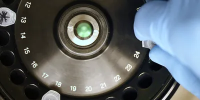
Live cell microscopy allows researchers to observe living cells in real time using time-lapse imaging. However, automated analysis remains difficult because cells change size, shape, and position over time, particularly during environmental stress or reproductive processes.

The research team developed an algorithm called FIEST (Frame Interpolation Enhanced Single-cell Tracking), which uses AI-based image processing to follow individual cells even when they undergo substantial morphological changes. The system was initially developed in yeast and later adapted to track bacteria, cancer cells, and human organoids, demonstrating broad applicability across biological research models.
According to project lead Orlando Arguello-Miranda, combining AI with microscopy significantly accelerated biological analysis.
“We have accelerated the analysis of living cells to a point we only could dream of four years ago. By combining generative AI and live cell microscopy, we are now able to track individual cells and microorganisms without interruption throughout their entire lives, including cell division, reproduction, and dormancy.”
Advantages for single-cell analysis workflows
AI cell tracking provides several benefits for laboratory operations and research workflows:
- Continuous monitoring across multiple cell generations
- Automated quantification of growth and behavior
- Reduced manual image analysis time
- Improved tracking accuracy compared with traditional methods
- Scalability for high-throughput imaging datasets
In one demonstration, the algorithm successfully tracked 632 individual cells in a complex microscopy dataset where previous tracking approaches failed, highlighting the potential for improved single-cell analysis reliability.
Expanding applications beyond basic research
As interest in the technology increased, researchers identified demand for scalable image analysis tools outside academic environments. To address this need, the team co-founded a platform that enables laboratories to upload microscopy images and apply AI-based tracking algorithms through a centralized system.
High-content microscopy experiments can generate thousands of images, creating analysis bottlenecks that slow research timelines. Automated AI cell tracking tools could help laboratories reduce these bottlenecks while improving data consistency across experiments.
The technology is also being adapted for agricultural and environmental applications, including rapid detection of plant pathogens using microscopy-based identification methods. AI analysis can identify distinctive spore shapes and determine whether pathogens are alive, offering advantages compared with DNA-based tests that detect genetic material but cannot confirm viability.
Future implications for laboratory research
Researchers are continuing to expand AI-enabled imaging approaches to study intracellular protein behavior and molecular interactions within living cells. Advances in AI cell tracking could enable scientists to monitor complex biochemical networks with higher temporal resolution, improving understanding of disease mechanisms, cellular signaling pathways, and organism development.
For laboratory managers and research organizations, improved AI-driven microscopy analysis may influence:
- Imaging infrastructure investments
- Data analysis workflows
- Automation strategies for high-throughput experiments
- Cross-disciplinary collaboration between computational and biological teams
As AI integration with microscopy technologies continues to evolve, laboratories may gain new capabilities to observe biological systems dynamically, rather than relying solely on static measurements, enabling more detailed insights into cellular processes and experimental outcomes.
This article was created with the assistance of Generative AI and has undergone editorial review before publishing.


