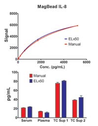
Figure 1. Comparison of ELx50 and manual vacuum washed cytokine calibration curve (top) and sample concentration determinations (bottom).
BioTek
Microplate-based multiplex assays, where microspheres are used as solid support matrices, provide simultaneous analyte analysis per well with lower background noise and reagent, consumable and sample costs compared to traditional ELISAs. Specific antibodies are conjugated to different identifiable beads made from a combination of plastics, ferrite, and other materials. A multi-analyte sample is added to beads in a microplate well, and will only bind to the beads if a specific affinity exists. Any unbound compounds remain in solution and are removed during subsequent wash steps. Non-magnetic bead-based assays are performed in filter-bottom microplates, where liquids are drawn out from the microplate bottom using a vacuum manifold, while magnetic bead-based assays use a strong magnet to immobilize the beads while liquid is removed. The beads are read on a flow cytometer, which identifies the specific bead type analyte, as well as quantitating the analyte.
Critical to optimal microsphere use is adequate washing. Manual washing increases risk of residual unbound material, bead-impregnated filter plates during vacuum processing or bead loss during vacuum processing or magnetic immobilization; each leading to inconsistent, unexpected, or incorrect results. A more effective alternative for vacuum- or magnetic immobilization-based multiplex assays is an automated microplate washer. Automating multiplex assay wash steps increases efficiency while reducing or eliminating operator error. Use of automated washers, such as BioTek’s ELx50™ microplate strip washer or ELx405™ microplate washer, allows for full washing parameter control, unattended operation and timely, reproducible and statistically valid measurements.
Here we describe automated vacuum and magnetic wash steps, including low residual volume, good bead recovery and optimal washing benefits using the ELx50 and ELx405. With the washers, we used polystyrene vacuum manifold-based and magnetic immobilization-based human cytokine multiplex assay kits from Bio-Rad and Invitrogen, respectively, and a LX100 reader from Luminex Corporation.
Multiplex assay workflow and performance
Figure 2. Comparison of ELx50 and manual magnetic-immobilization washed cytokine calibration curve (top) and sample concentration determinations (bottom).
BioTek
To assess the multiplex assay performances using both wash methods, assay kit bead mix was added to a filter plate (vacuum workflow) or flat-bottom (magnetic work-flow) microplate after conditioning, and the beads were washed using assay wash buffer. Multiplex standards containing 10 different analytes were generated by serial dilution of a reconstituted human cytokine mix standard. After reconstitution, each standard and sample were pipetted into microplate wells containing multiplex bead mixes and incubated per assay instructions. The plates were then vacuum aspirated or magnetically immobilized and visually inspected for completeness, followed by two dispense and vacuum or magnetic immobilization aspirations. After washing, detection or secondary antibody reagent was added and incubated per assay instructions. The beads were again washed followed by a SAPE reagent addition. After agitated incubation for reporter tag binding, the plates were again washed as instructed. Samples and standards were then resuspended in assay buffer and read on the Luminex reader using assay kit parameters. Each assay was also washed manually for comparison purposes.
Per Figure 1 (top), a typical analyte standard curve was generated by plotting the median fluorescent intensity (MFI) signal against concentration. These curves, generated for manual and automated (ELx50) vacuumbased washing methods, show a high degree of similarity. Additionally, calculated low, medium and high sample concentrations (Figure 1, bottom) also show strong correlation between manual and automated wash methods.
The similarity in manual versus analyte standard curve generation and sample concentration determination also holds true for magnetic-based washing methods (Figure 2, top and bottom).
Figure 3. Standard curves for the magbead multiplex human cytokine panel using ELx405 microplate washer.
BioTek
Finally, using known analyte concentrations, standard curves were generated for each cytokine by plotting the MFI signal against concentration. These standard curves are then interpolated to determine the unknown sample concentrations. Again, efficient washing to remove non-specific antibody binding is critical for dependable results. Per Figure 3, very reliable data was generated using the magnetic bead-based multiplex assay with an automated (ELx405) wash method in 96-well microplate format. This data was consistent from run-to-run and day-to-day. These standard curves may be used to calculate unknown sample concentrations with high confidence.
Conclusions
Multiplex assay methods provide researchers with more information and higher sensitivity in less time and with fewer materials than traditional ELISAs. Automating the wash steps in vacuum and magnetic multiplex methods provides improved repeatability, ease-of-use and convenience compared to manual methods.