 Scanning avoids physically taking the bee’s brain apart.Image courtesy of the University of GuelphDetailed exploration of tiny insect brains has become much easier using new methods for imaging and 3D image reconstruction developed by a team of scientists, including a University of Guelph professor.
In a study published Feb. 24 in Scientific Reports, the team discusses use of micro-computed tomography (micro-CT) to examine bumblebee brains.
The researchers used this X-ray imaging to produce hundreds of image slices that can be re-constructed by a standard laptop computer into a high resolution 3D model, all without destroying the sample.
Prof. Nigel Raine, Rebanks Family Chair in Pollinator Conservation in the School of Environmental Sciences, worked with researchers at Imperial College and the Natural History Museum in London on the new method, which images the bees’ heads without mechanical trauma.
“This metholodogy provides a much more accurate and realistic image of the shape, size and interactions of soft tissues in the very small brains of insects,” he said.
Related Article: World-Class Honey Bee Facility Reveals Insights into Workings of Human Brain
A bee brain contains approximately 1 million nerve cells, which equates to 0.00001 per cent of the number found in the human brain.
The new paper assesses staining techniques to enhance brain image contrast and the use of freely available software to reconstruct the shape of brain structure from high-resolution scans on a normal laptop computer.
“Our approach increases the accessibility of this technique to a wider audience of researchers,” Raine said.
He said this technology will help researchers better understand how variations in bees’ brains affect their behavior.
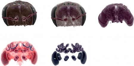 Researchers used CT scanning to image bee brains.Images courtesy of the University of Guelph“Variation among individual bees or bee colonies in their learning abilities has important consequences for essential behaviors, such as the ability to collect nectar and pollen efficiently,” he said.
“We’ve also been looking at how tiny-brained bumblebees find practical solutions to challenging routing problems. Understanding how comparatively simple brains can find functional solutions to complex problems may be very important in allowing us to develop smarter and simpler ways to do the same.”
Raine said researchers need to know more about what affects the behavior of insects, including how bee brain structures respond to environmental stresses such as disease, poor nutrition, or pesticide exposure.
For example, he said, “small changes in the structure and shape of brain structures like the mushroom bodies resulting from environmental stress could have big implications on bee behavior. This approach to imaging will help us to investigate these possible effects and the consequences they could have for bee health and the important pollination services they provide to crops and wild plants.”


