Physiologically relevant testing platforms
For example, whereas most somatic cells exist in close proximity to other cells and support matrices, bio-production cells grow in suspension.
By contrast, cells used in research and development in cell-based assays are cultured as closely as possible to their original state, which whenever feasible means three-dimensional culturing.
Three-dimensional (3-D) culturing preserves cell-cell interactions that are essential for recapitulating the cells’ natural milieu, thereby generating more realistic data for efficacy and toxicity studies of drugs, cosmetics, foods, industrial chemicals, household products, and other items. Increasingly, 3-D cell-based assays are replacing research animals.
Coculture's the thing
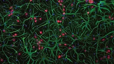
For Danilo Tagle, PhD, associate director for special initiatives at the National Center for Advancing Translational Sciences (NCATS), part of NIH, coculture is the defining characteristic of 3-D culture. “Two-dimensional cultures consist of relatively homogeneous cells, whereas 3-D cultures are heterogeneous in terms of cell type, morphology, and physical properties,” Tagle says.

The NIH’s tissue chip program’s approach is unique in that it begins with induced pluripotent stem cells (IPSCs) derived from adult tissues and reprogrammed into pluripotent cells that differentiate into many specific cell types, including endothelial cells that make up blood vessels and neurons that turn into nerves. When the different cell types combine within the proper niche, they recognize each other and self-assemble into structures analogous to those found in the body.
“The presentation of microvasculature in tissue chips is essential,” Tagle says. The tiny channels are capable of introducing test materials to cells and removing waste products in a physiologically relevant manner. Tagle’s lab is currently attempting to insert innervation to reproduce physiologic responses to biomechanical stresses.
Another unique aspect of the NIH tissue chips is they are monoclonal—all cellular elements derive from a single human, so all cells are genetically identical. Differences in genomic composition could prevent the happy coexistence of cells in proximity, or cause them to behave erratically when challenged by a stimulus. In a worst-case scenario, tissue components could initiate immunologic responses against each other. Monoclonal chips are possible thanks to advances in IPSC technology.
The NIH group is not quite at the point where it can produce all relevant cells from stem cells. “That goal remains a work in progress,” Tagle says.
“We can still learn many things from 2-D cell culture,” Tagle says, “but if we’re trying to be scientifically exact, we must use 3-D constructs that include microfabricated or bioengineered scaffolds rather than standard ‘culture ware,’ complete with the microfluidic flow and re-creation of biomechanical responses to stress that tissues exhibit in their native environment.”
For example, lung tissue stretching in response to breathing induces lung cells to produce surfactants that may enhance or interfere with the effectiveness or toxicology of drugs. Lung cells cultured in 3-D do indeed stretch and secrete high levels of surfactant. Similarly, hepatocytes cultured under physiologically relevant conditions produce more albumin and urea.
The complexity of Tagle’s group’s projects is breathtaking. For example, there are now integrated heart-liver-vascular systems; circulatory systems integrated with muscle; human cardiopulmonary systems “on a chip”; all-human microphysical models for cancer metastasis therapy; and human, IPSC, and embryonic stem cell-based models for predictive neural toxicity and teratogenicity.
The game-changing and highly lucrative field of regenerative medicine has been a boon to the organ-on-chip industry that now serves as a stepping stone between basic science and final products.
Not all organizations necessarily focus on regenerative medicine, however. Tagle’s group’s main objective is producing the best organ chips available for R&D. Yet Tagle recognizes what he calls “two-way traffic,” a symbiosis between organ chips and manmade organs. Several of Tagle’s group’s members have regenerative medicine experience. “The lessons we’re learning on how cells mature, grow, assemble, and relate to one another undoubtedly can benefit regenerative medicine and vice versa.”
Keeping cells happy
Aside from greater complexity, what differentiates 3-D from 2-D culture is that cells grown in 3-D are healthier, live longer, and are more productive. “Not having a lot of dead cells around is critical for cultures that eventually are infused or implanted into patients,” says Alex Sim, president, AMSBIO (Cambridge, MA). AMS has developed a method for removing dead cells from cultures through a technique that identifies surface markers found on dead cells.
Culturing within hydrogels or extracellular matrix is suitable for monocultures, but arguably the greatest benefit of 3-D is the ability to coculture two or more cell types. Examples include a coculture of fibroblasts and cancer cells to observe interactions related to tumor aggressiveness, or a triculture model incorporating vascular and stromal cells with breast cancer tumor spheroids mimicking the environment, cellular architecture, and behavior of actual tumors. “You can’t otherwise get that information without using an animal,” Sim says.
Organoids are composed of 3-D cultures formed from stem cells that have been induced into specific tissue lineages that subsequently self-assemble into meaningful structures. Through organoids, scientists can create monoclonal tissues for the variety of assays open to conventional 3-D culture. Ultimately, one could apply them to personalized or regenerative medicine.
Organoids cultured from a single patient could be employed as live-cell screens for personalizing cancer treatment.
Relevant, but not 3-D
“The discussion around 2-D or 3-D culture comes down to what is more realistic, which format better approximates physiologic conditions,” says Katherine Cook, global marketing manager at Hepregen (Medford, MA).
Hepregen’s HepatoPac products, for example, are more complex than 2-D cultures, but do not strictly qualify as 3-D. HepatoPac is an in vitro liver model designed for both short- and long-term preclinical drug toxicology as well as for efficacy studies. Micro- patterning of hepatocyte islands with supportive stromal cells replicates the physiological microenvironment of the liver, promoting hepatocyte health and supporting metabolic activity for more than four weeks.
“Hepatocytes are normally very difficult to grow in culture,” Cook explains. “In the liver, they are surrounded by a host of other cell types with which they exchange chemical signals critical to maintaining hepatic function.”
Hepatocyte island technology is based on research by Hepregen’s co-founder and scientific adviser (and current MIT professor), Sangeeta Bhatia, MD, PhD. Employing technology from the semiconductor industry, Bhatia etched dozens of different physical matrix patterns into culture ware before discovering the architecture and ratio of multicellular interactions that best supported the coculture of hepatocytes and stromal cells. She eventually settled on micro-patterns of a specific diameter, with hepatocytes adhered to extracellular matrix-coated islands and supportive, stromal cells adhered to the space in between.
In the liver, hepatocytes are specifically arranged (polarized) in such a way that they can take up compounds from the blood while secreting metabolized compounds into the bile. As such, hepatocyte cells are arranged in plates that are at most one to two cells thick. Cells in HepatoPac are similarly one to two cells thick and in contact with media and internal networks of bile capillaries.
Self-assembling aggregates
Not all 3-D cultures require a specific growth matrix. Spheroids are self-assembled multicellular aggregates that produce their own extracellular matrix, and encompass the complex cell-matrix and cell-cell interactions that mimic functional in vivo tissues. Specifically, spheroids mimic avascular tumors with inherent metabolic (oxygen, carbon dioxide, nutrients, wastes) and proliferative gradients, thus they serve as tumor models, providing reliable and meaningful therapeutic results compared to 2-D culture tests.
Meanwhile, 3-D spheroids have historically been difficult to produce and not well suited to high-throughput screening. The emergence of hanging drop plates (HDPs) greatly facilitated spheroid production. HDPs are available from several distributors as well as the OEM vendors InSphero (the GravityPLUS™ product) and 3D Biomatrix (the Perfecta3D® product).
HDPs facilitate controllable, consistent formation of 3-D spheroids. A drop of cell suspension is pipetted into the top of each well using common manual or automated liquid-handling equipment, and the plate geometry causes the suspension to hang stably below the well where the cells aggregate into a spheroid over one to five days, depending on the cell type. The user can control the spheroid diameter through the type and number of cells dispensed into each well. Access holes at the top of each well allow for media exchange and the addition of compounds, reagents, or additional cells to establish patterned cocultures. Without contact with any surfaces or matrices, one spheroid forms per well.
Spheroids are analyzed using absorbance, fluorescence, and luminescence assays using a plate reader. Microscopic imaging is also possible. The platform also offers simplified liquid-handling procedures and compatibility with HTS instruments and high-content imagers.
“The 2-D tissue culture models lack realistic complexity, while animal models are expensive, time-consuming, and too frequently fail to reflect human biology,” comments 3D Biomatrix CEO Laura Schrader. Most important is preserving cell-cell contacts, which Schrader believes is facilitated by eliminating artificial substrates coming into contact with cells. “This allows development of proliferative zones and gradients of gases, nutrients, and wastes.”
Like other 3-D techniques, HDPs are amenable to most cell types, including primary cells, immortalized cell lines, and stem cells. 3D Biomatrix estimates that half of all cancer cell lines form spheroids spontaneously in HDPs. For those that do not, addition of exogenous extracellular matrix or stromal cells will generally promote spheroid formation.
No single 3-D culture method serves all end uses. And despite its advantages, 3-D culture is not always the most appropriate culture method for every application. Most cultures today are, in fact, of the 2-D or suspension variety, many of which are relevant enough to provide useful data. “But organs are not composed of monolayers. If true physiological relevance is your goal, you should at least be thinking of 3-D culture,” observes Sim.


