Making Small Things Visible
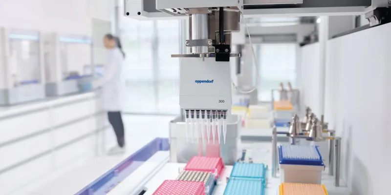
 BX63 / Automated Fluorescence Microscope Olympus / www.olympus.com Microscope-based imaging used to be the domain of core facilities and dedicated operator-directors. Today, imaging has become routine—not quite to the “technician” level for all techniques but moving in that direction. Motorized stages and other automation tools are enabling unattended imaging of multiple samples. Confocal systems that once required a dark room now sit on fully lighted laboratory benches.
But as microscopy advances and the “heart cut” methods become more democratized and accessible, the leading edge becomes more complex in terms of science and instrumentation. Together these advances “change how people address questions in biology,” says Brendan Brinkman, senior product manager for laser scanning confocal microscopes at Olympus (Center Valley, PA). “The same individuals who might have used a benchtop fluorescence microscope several years ago now have access to imaging systems that were the exclusive tools of research labs.”
Microscopic imaging has come a long way during the past decade, Brinkman adds. “Confocal microscopy has become established to the point where people view it as routine.” Brinkman cites multiphoton imaging as another “routine” technique, particularly for visualizing in vivo processes. The National Institutes of Health BRAIN (Brain Research through Advancing Innovative Neurotechnologies) Initiative relies heavily on these two methods. Interest in fixed tissue imaging has not slackened either, according to Brinkman.
Pittcon 2014 saw the debuts or formal introductions of at least 25 microscopes, most of them suitable for imaging applications; for example:
- EDAX demonstrated its EBSD (electron backscatter diffraction) for SEM. With a focus on analysis of materials with crystalline structures, EBSD allows users to analyze orientation, grain morphology, material deformation, and distinct crystal phases.
- Carl Zeiss introduced the EVO SEM for materials characterization and quality assurance.
- Thermo Fisher showed the DRX™xi Raman microscope, described below.
- Olympus unveiled its BX63 imaging microscope with full motorized control, a unique focusing mechanism, and cellSens Dimension software for cell imaging.
Microscopy is an ideal imaging platform, because it operates in size domains that are generally invisible to the naked eye. The application of spectroscopy (e.g., Raman, infrared, ultraviolet, fluorescence) to microscopy further expands microscopic visualization of chemical components that appear identical with visible light microscopy. Chemical mapping of materials, tissues, and cells has become the leading edge of microscale imaging.
To illustrate the importance of microscope-based imaging across size domains of many orders of magnitude, consider that a fair amount of diagnostic imaging today occurs not on arms, legs, and livers or on tissues or cells but at the molecular level—for example, fluorescence in situ hybridization (FISH), which is described in greater detail below.
Imaging by mass Spectrometry
Microscopy is not the only imaging platform suitable for microscale samples and events. The ability to discriminate on the basis of molecular weight is what confers similarly useful imaging capabilities on mass spectrometry. Although MS is not microscopy, techniques that combine sampling of very small regions on samples, gentle ionization, and software that renders mass data and physical coordinates into composition maps have pushed MS to the forefront of advanced imaging technologies, albeit one that involves greater capital expenses than light microscopy.
Because of its mild ionization mechanism, MALDI (matrix-assisted laser desorption ionization) has been the mass spectrometry imaging technique of choice for years. All major MS companies sell MALDI systems. Both commercial and public domain image software exists as well.
MALDI imaging takes place on thin tissue samples where the mass of one or more target molecules is known. With assistance from software, MS creates 2-D distribution images of drugs, natural products, or metabolites along a tissue cross-section. The sample is first treated with a matrix solution that assists in volatilization and ionization. Then a laser focused on tiny “pixels” vaporizes a bit of sample, which is swept into the spectrometer. Software then creates a 2-D distribution image for the molecule of interest.
MALDI may be combined with other imaging methods— for example, magnetic resonance—or by taking successive tissue slices, in order to create 3-D images.
MS imaging of large, complex molecules would not be possible without gentle ionization. Otherwise, ionization would destroy or disrupt proteins and other large, labile molecules. An emerging MS imaging platform, LAESI (laser-assisted electrospray ionization), is based on mild electrospray ionization (ESI) but has several advantages over MALDI.
For one, LAESI essentially eliminates matrix addition and, more significantly, sample prep. “Matrix addition can create artifacts that can be bypassed by eliminating sample prep entirely,” observes Haddon Goodman, LAESI platform marketing manager at Protea Biosciences (Morgantown, WV).
Unlike imaging methods that require contrast agents or labeling with radioactive compounds and sophisticated image acquisition instrumentation, “molecular imaging” exemplified by MALDI and LAESI relies only on the masses of target analytes, which are “filtered” by the spectrometer. LAESI can create multiple chemical maps of a surface simply by filtering a panel of masses.
The critical difference between molecular imaging and, say, MRI is that the latter usually takes place in living organisms, whereas MS methods cannot. “You need a slice of tissue,” Goodman says.
Where both LAESI and MALDI create 3-D images through sectioning, LAESI alone is capable of tunneling through samples from point to point. This technique has been demonstrated for plant tissues, where cell walls maintain structural integrity. Goodman says his company is working on a similar method for animal tissue.
LAESI’s most interesting imaging strength has to do with rapid screening of surfaces without sample prep. In addition to plant and animal tissue, LAESI is capable of scanning surfaces of cultured cells in plates or Petri dishes. Researchers are using this technique to investigate biomarkers and the effects of drug treatments among microbial colonies, to identify natural products for drug discovery, and to visualize downregulation and upregulation of genes. “You can tell if transfection worked by imaging regions where proteins encoded by that gene are expressed,” Goodman says.
LAESI enhances the capabilities of conventional medical imaging techniques as well. In September 2013, Protea entered a collaborative research agreement with Virginia Commonwealth University’s Center for Molecular Imaging. The collaboration will combine LAESI with the university’s in vivo molecular PET, SPECT, and MRI/MRS medical imaging capabilities to investigate the molecular basis of cancer, Alzheimer’s, and other human diseases.
MS has brought about changes in imaging-based diagnosis of tissues that microscope-based imaging could not. For example, researchers from Imperial College, London, use MS data from a tissue sample to create an image of the sample’s chemical composition. The technique maps chemical components of interest and may help medical researchers differentiate tissues that appear similar under ordinary light microscopy. For example, mass spectral imaging could indicate differences between cancer subtypes.
This advance pushes the limits of conventional pathology/ histology, which over the past 50 years have relied on dyes, stains, and light microscopy and take many days. MS imaging will enable chemical composition data mapping but, more important, make this type of data accessible to clinicians and researchers. Dr. Zoltan Takats at Imperial College calls MS imaging a paradigm changer. “Instead of defining tissue types by their structure, we can define them by their chemical composition. This method is independent of the user—it’s based on numerical data rather than a specialist’s eyes—and it can tell you much more in one test than histology can show in many tests.”
Raman microscopy— Unveiling the invisible
Raman microscopy complements optical microscopy and other imaging tools by providing chemical and morphological analysis at the same microscopic level of detail. Because Raman measures molecular bond vibrations— essentially chemical fingerprints—users obtain information that is inaccessible via light microscopy.
“Raman reveals otherwise invisible information to increase understanding and solve problems in a wide range of materials, from pharmaceutical tablets to graphene monolayers to minerals,” says Scot Ellis, marketing manager for Raman spectroscopy and microscopy at Thermo Fisher Scientific. “Raman is becoming an essential competitive tool in academic and industrial research.”
 Raman Imaging Microscope / DXR™xi / Thermo Fisher Scientific www.thermoscientific.com Because it resolves objects at submicron levels, Raman is used for a wide variety of scientific problems, from verifying chemical composition and finding/identifying contaminants in products to optimizing production processes.
According to Ellis, the Thermo Scientific™ DXR™xi Raman imaging microscope is a new approach to Raman imaging. “It differs from traditional systems by working at the problem level, that is, an entire chemical image, rather than depending on a user to work with individual point spectra to build an image up.”
The DRXxi rasters images by collecting spectral data at a very high rate, statistically processing and interpreting behind the scenes and displaying results during collection. Color staining with chemical or morphological information reveals “many dimensions of data,” Ellis says. “Chemical identification and distribution, physical information such as materials strain, and relative concentration information all are conveyed visually.”
The DXRxi supplements many microscopic techniques that provide visual or topographical information and that often require expertise, subjective interpretation, or additional measurements that must be correlated to the microscopic image to be meaningful.
“More important, the DXRxi replaces traditional Raman microscopes by operating in an entirely different way,” Ellis says, by delivering meaningful chemical images nearly instantaneously. “It’s really microscopy powered by spectroscopy, whereas historically, systems have worked the other way around.”
A twist on fluorescence
Through its use of fluorescently labeled tags, fluorescence microscopy provides a means of targeting specific structures inside cells.
Fluorescence in situ hybridization (FISH) is a technique that images abnormal genes that are indicative of disease. In cancer disease management, for example, FISH detects targeted DNA abnormalities and is widely used for diagnosis, prognosis, and treatment selection.
Microscope manufacturers are particularly interested in FISH because it uses fluorescence to image very small objects. In 2013 Nikon Instruments entered an agreement with Cancer Genetics (Rutherford, NJ) to distribute FISH DNA probes for oncology. The deal brings together the two ingredients necessary for DNA-level diagnostic imaging: fluorescent FISH probes (from Cancer Genetics) and a suitable microscopy-imaging platform (from Nikon). In this instance, the genetic tests will take advantage of a technique Nikon has developed for performing simultaneous, multicolor FISH testing and complex image analysis.
While the technology behind FISH is not new, fluorescence- based techniques are rapidly evolving, together with (and because of) instrumentations; for example, multicolor FISH. A variation on this theme is two-photon or multiphoton fluorescence.
Bruker’s (Billerica, MA) acquisition of Prairie Technologies last autumn afforded Bruker an entry into multiphoton fluorescence microscopy, a microscopy imaging technology in which Prairie was a pioneer. In traditional fluorescence the excitation energy is shorter in wavelength (and higher in energy) than the emitted fluorescence. In multiphoton, two or more photons of longer wavelength (lower energy) strike the target simultaneously, producing an emission of higher energy than the excitation does.
Bruker also gained entry into confocal microscopy, a high-resolution, high-contrast imaging technique that employs point illumination and pinhole masking to eliminate out-of-focus light.
Although the combined excitation energy of the two photons may be the same or higher than in single-photon fluorescence, the impact on living organisms is gentler. Think of being hit by two tennis balls traveling at 25 miles per hour versus one ball at 50 miles per hour. Consequently, dual-photon fluorescence is ideal for imaging live cells that are able to withstand just so much excitation. Cells survive longer, so analysts can extract more information from them.
Multiphoton fluorescence enables exotic-sounding experiments such as uncaging, optogenetics, simultaneous electrophysiology, and photoactivation techniques such as photostimulation and photablation. All these involve selective, targeted interaction between light and matter, usually with cells, resulting in perturbations detected by image-capture and rendering elements.
Jeff Stuckey, PhD, product marketing manager for fluorescence microscopy at Bruker Nano, formerly of Prairie Technologies, explains that photoactivation does not actually move sample constituents in space. “For that you need laser tweezers, which we don’t do. Instead, we activate molecules using laser light.” In caging experiments, for example, lasers cause the release of neurotransmitters from nerve cells. In another form of photoactivation, optogenetics, neurons are stimulated to mimic naturally occurring neuronal activity, which allows investigators to image neuronal connections and activity.
“[Because] fluorescence occurs only where photons arrive simultaneously, fluorescence is limited to tiny, well-defined regions,” Stuckey explains. The analogous confocal imaging technique results in diffuse fluorescence above and below the desired location, resulting in photobleaching, which contributes to out-of-focus light.
Another advantage of two-photon fluorescence excitation involves its use of infrared lasers, which penetrate more deeply than visible light into tissue. Where visible wavelengths image to a depth of about 300 microns, two-photon systems reach five times as deep, to 1.5 millimeters. Through the use of clearing agents that render opaque tissues clear, scientists have achieved imaging depths of eight millimeters.
Don’t forget industrial imaging
Industrial imaging includes nondestructive macroscale techniques for visualizing defects, composition, and other relevant features of materials or structures. In keeping with the spirit of this article, we will limit the discussion to microscope-based imaging, for which several dozen optical and nonoptical techniques exist.
FEI (Hillsboro, OR) focuses mostly on scanning electron microscopy (SEM), dual-beam focused ion beam/ SEM (FIB/SEM), and transmission electron microscopy (TEM). Through its acquisition of Germany-based Till Photonics, FEI has recently invested in light microscopy as well. In early 2014 FEI announced that it had acquired Australian firm Lithicon, which provides pore-scale micro-computed tomography (μCT, or microCT) equipment to oil and gas companies.
A significant segment of FEI’s business involves atomicscale imaging, which encompasses molecules as well as atoms. Relevant instrumentation includes TEM and scanning transmission electron microscopy (S/TEM). TEM uses a focused beam smaller than the atoms themselves to sample along a group of atoms. Bert Freitag, director of product marketing for FEI’s materials science business unit, compares STEM to a “slide projector” that illuminates a larger area to collect data and transmit it to a CCD camera.
 Ultrafast Electron Microscope / Tecnai™ Femto / FEI / www.fei.com TEM in particular has evolved as a cutting-edge technique for atomic imaging, but it was limited to visualizing atoms in “black and white.” It is now possible through advanced imaging to assign “colors” to atoms based on spectroscopic properties. “This allows us to visualize chemical information,” Freitag says. The two techniques employed by FEI are energy-dispersive X-ray analysis (EDS) and electron energy loss spectroscopy (EELS).
Another advanced STEM technique, differential phase contrast, allows measurement of electric fields between atoms, which are directly linked to the physical properties we experience in the macroscopic world. The technique also visualizes electric fields between atoms, which are significant in polar ceramics such as gallium nitride, used to manufacture light-emitting diodes. “These fields are directly linked to light-emitting properties, so understanding them can help in the design or improvement of these materials,” Freitag says. “Information at the atomic scale provides many significant insights into the macroscopic properties of materials.” The observation of variation of atomic distances can provide information about strain, for example, in silicon lattices of nanodevices, which directly relates to electron mobility and faster-switching transistors.
FEI has continued its innovation in visualizing ultrasmall, ultrafast events with its late 2013 introduction of the Tecnai Femto ultrafast electron microscope. The device allows observation of events occurring at the atomic and molecular scale, within time frames of femtoseconds (10-15 second). These include the absorption of light energy and its transformation into heat, mechanical changes through photoactuation, and crystallization. Tecnai Femto is the first system to commercialize ultrafast electron microscopy technology pioneered by Nobel laureate Prof. Ahmed Zewail at the California Institute of Technology.
David Flannigan, PhD, a former Zewail lab member and now assistant professor of materials science at the University of Minnesota, has noted that “over the last decade microscope manufacturers like FEI have developed instruments that have made observations of objects as small as individual atoms relatively routine. Ultrafast electron microscopy now gives us a powerful tool to look at the movements and changes that occur at this scale. Because the distances are so small, the time scale is also condensed—it doesn't take very long to travel a nanometer or two. Using single-electron pulses, we have measured changes over time periods as short as tens of femtoseconds— those are millionths of a billionth of a second.”