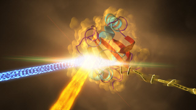
 This illustration depicts an experiment at SLAC that revealed how a protein from photosynthetic bacteria changes shape in response to light in less than a trillionth of a second. Samples of the crystallized protein (right), called photoactive yellow protein, or PYP, were struck by an optical laser beam (blue light coming from left) that triggers shape changes in the protein. These were then probed with a powerful X-ray beam (fiery beam from bottom left) from SLAC’s LCLS.Image credit: SLAC National Accelerator Laboratory
MENLO PARK, CALIF. — A number of important biological processes, such as photosynthesis and vision, depend on light. But it’s hard to capture responses of biomolecules to light because they happen almost instantaneously.
Now, researchers have made a giant leap forward in taking snapshots of these ultrafast reactions in a bacterial light sensor. Using the world’s most powerful X-ray laser at the Department of Energy’s SLAC National Accelerator Laboratory, they were able to see atomic motions as fast as 100 quadrillionths of a second—1,000 times faster than ever before.
Further, “We’re the first to succeed in taking real-time snapshots of an ultrafast structure transition in a protein, in which a molecule excited by light relaxes by rearranging its structure in what is known as trans-to-cisisomerization,” says the study’s principal investigator, Marius Schmidt from the University of Wisconsin-Milwaukee.
The technique could widely benefit studies of light-driven, ultrafast atomic motions. For example, it could reveal:
- How visual pigments in the human eye respond to light, and how absorbing too much of it damages them.
- How photosynthetic organisms turn light into chemical energy—a process that could serve as a model for the development of new energy technologies.
- How atomic structures respond to light pulses of different shape and duration—an important first step toward controlling chemical reactions with light.
“The new data show for the first time how the bacterial sensor reacts immediately after it absorbs light,” says Andy Aquila, a researcher at SLAC’s Linac Coherent Light Source (LCLS), a DOE Office of Science User Facility. “The initial response, which is almost instantaneous, is absolutely crucial because it creates a ripple effect in the protein, setting the stage for its biological function. Only LCLS’s X-ray pulses are bright enough and short enough to capture biological processes on this ultrafast timescale.” The results were published May 5 in Science.


High-speed X-ray Camera Reveals Extremely Fast Biology
The team looked at the light-sensitive part of a protein called “photoactive yellow protein,” or PYP. It functions as an “eye” in purple bacteria, helping them sense blue light and stay away from light that is too energetic and potentially harmful.
The researchers had already studied light-induced structural changes in PYP at LCLS, revealing atomic motions as fast as 10 billionths of a second. By tweaking their experiment, they were now able to improve their speed limit 100,000 times and capture reactions in the protein that are 1,000 times faster than any seen in an X-ray experiment before.
Both studies followed a very similar approach: At LCLS, the team sent a stream of tiny PYP crystals into a sample chamber. There, each crystal was struck by a flash of optical laser light and then an X-ray pulse, which took an image of the protein’s structural response to the light. By varying the time between the two pulses, scientists were able to see how the protein morphed over time.
Since LCLS’s X-ray pulses are extremely short, lasting only a few quadrillionths of a second, they can in principle probe processes on that very timescale—but only if the optical laser also matches the tremendous speed. For the new experiment, the team replaced the old optical laser with a new one whose pulses were 100 quadrillionths of a second long—100,000 times shorter than before and much closer to the X-ray pulse length.
The researchers also applied better timing tools to measure the relative arrival time between the optical and X-ray laser pulses, enhancing the ability to precisely track ultrafast events.
“These improvements allowed us to see what no one has ever directly seen before,” Schmidt says.
Other institutions involved in the study were: Center for Free-Electron Laser Science/Deutsches Elektronen-Synchrotron, Germany; Imperial College, UK; University of Jyväskylä, Finland; Arizona State University; Max Planck Institute for Structure and Dynamics of Matter, Germany; State University of New York at Buffalo; University of Chicago; Lawrence Livermore National Laboratory; and University of Hamburg, Germany. Funding sources included: National Science Foundation; National Institutes of Health; Helmholtz Association; German Federal Ministry of Education and Research; Engineering and Physical Sciences Research Council; Academy of Finland; and the European Union.