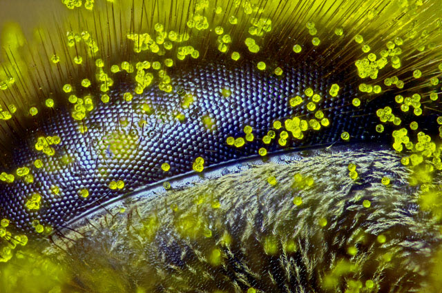
 First place: Eye of a honey bee (Apis mellifera) covered in dandelion pollen (120x).Photo credit: Ralph Claus GrimmNikon Instruments Inc. today (Oct. 14) brings the world eye-to-eye with a honey bee, awarding first place of the annual Nikon Small World Photomicrography Competition to Australian Ralph Grimm for his incredible close-up image of a bee eye covered in dandelion pollen grains. Grimm’s winning image is not only visually striking, but brings to light just how little is understood about how these incredible insects see the world.
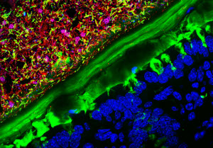 2nd Place: Mouse colon colonized with human microbiota (63x)Photo credit: Kristen Earle, Gabriel Billings, KC Huang & Justin Sonnenburg, Stanford University School of MedicineIn addition to Grimm, Nikon recognized over 77 other winners of the 2015 Small World competition, including a ranked Top Twenty, 12 Honorable Mentions and 56 Images of Distinction. With submissions spanning 83 countries, competition was tough. Judges selected winners that displayed not only artistic quality but exceptional scientific technique.
“Each year we are blown away by the incredible quality and quantity of microscopic images submitted from all over the world, from scientists, artists, and photomicrographers of all levels and backgrounds. This year was certainly no exception,” said Eric Flem, Communications Manager, Nikon Instruments. “Judges had their work cut out for them in narrowing down from such a rich pool of applicants, and we are so pleased with the results. Each of these winning images exhibits the exemplary technique, scientific discipline and artistry for which Nikon Small World is known.”
Judges were particularly impressed by the masterful technique Grimm employed to capture this image stack, which included over four hours of careful work to mount the eye, set the focus increments, properly illuminate the subject and avoid peripheral smudging during the stacking process. The resulting image is a testament to Grimm’s painstaking efforts.
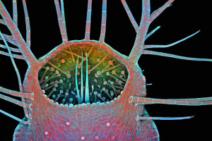 3rd Place: Intake of a humped bladderwort (Utricularia gibba), a freshwater carnivorous plant (100x)Photo credit: Dr. Igor Siwanowicz, Howard Hughes Medical Institute (HHMI)The story behind the image is also touching. As a high school teacher, self-taught photomicrographer and former beekeeper, the subject matter is near and dear to Grimm’s heart. While bee colonies continue to dwindle worldwide, Grimm hopes his image can serve as a voice for this rapidly disappearing insect that plays such a critical function in pollinating the world’s crops.
“In a way I feel as though this gives us a glimpse of the world through the eye of a bee,” says Grimm. “It’s a subject of great sculptural beauty, but also a warning- that we should stay connected to our planet, listen to the little creatures like bees, and find a way to protect the earth that we all call home.”
Ralph Grimm now joins the ranks of 37 other photomicrographers, artists and scientists from all over the world who have taken the top prize. This year’s competition received over 2,000 entries from more than 83 countries around the world.
Top Five Images:
1. Ralph Claus Grimm, Eye of a honey bee (Apis mellifera) covered in dandelion pollen
2. Kristen Earle, Gabriel Billings, KC Huang & Justin Sonnenburg, Mouse colon colonized with human microbiota
3. Dr. Igor Siwanowicz, Intake of a humped bladderwort (Utricularia gibba), a freshwater carnivorous plant
4. Daniel H. Miller & Ethan S. Sokol, Lab-grown human mammary gland organoid
5. Dr. Giorgio Seano & Dr. Rakesh K. Jain, Live imaging of perfused vasculature in a mouse brain with glioblastoma
The exceptional panel of judges who select the winning images has a tradition of including some of the most distinguished names working in the scientific community and science journalism today. The 2015 panel includes:
• Jacqueline Howard, Science Associate Editor, Huffington Post
• Ernie Mastroianni, Photo Editor, Discover Magazine
• Dr. Tim Mitchison, Vice-chair of the Department, and Co-chair of the PhD program in Systems Biology, Harvard Faculty and Sciences in Cambridge
• Dr. Hari Shroff, Chief and Tenure-track investigator, Section on High-Resolution Optical Imaging, at the National Institute of Biomedical Imaging and Bioengineering (NIBIB)
Top images from the 2015 Nikon Small World Competition will be exhibited in a full-color calendar and through a national museum tour.
Nikon Small World In Motion
For the first time, Nikon will also unveil the winners of its sister competition, Nikon Small World in Motion, in December 2015. Celebrating and showcasing the best of science and art under the microscope in the form of video, the 2015 Small World in Motion winners will be revealed on December 2, 2015 on www.nikonsmallworld.com.
See the 2015 winners


