
Two heads are better than one, as the saying goes, and sometimes two instruments, ingeniously recombined, can accomplish feats that neither could have done on its own.
Such is the case with a hybrid microscope, born at the Marine Biological Laboratory (MBL), that for the first time allows scientists to simultaneously image the full 3D orientation and position of an ensemble of molecules, such as labeled proteins inside cells. The research is published this week in Proceedings of the National Academy of Sciences.
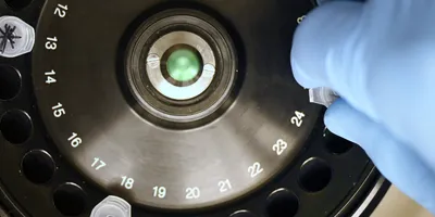
The microscope combines polarized fluorescence technology, a valuable tool for measuring the orientation of molecules, with a dual-view light sheet microscope (diSPIM), which excels at imaging along the depth (axial) axis of a sample.
This scope can have powerful applications. For example, proteins change their 3D orientation, typically in response to their environment, which allows them to interact with other molecules to carry out their functions.
"Using this instrument, 3D protein orientation changes can be recorded," said first author Talon Chandler of CZ Biohub San Francisco, a former University of Chicago graduate student who conducted this research partly at MBL. "There's real biology that might be hidden to you from just a position change of a molecule alone," he said.

Imaging the molecules in the spindle of a dividing cell -- a longstanding challenge at MBL and elsewhere -- is another example.
"With traditional microscopy, including polarized light, you can study the spindle quite nicely if it's in the plane perpendicular to the viewing direction. As soon as the plane is tilted, the readout becomes ambiguous," said co-author Rudolf Oldenbourg, a senior scientist at MBL. This new instrument allows one to "correct" for tilt and still capture the 3D orientation and position of the spindle molecules (microtubules).
The team hopes to make their system faster so that they can observe how the position and orientation of structures in live samples change over time. They also hope development of future fluorescent probes will enable researchers to use their system to image a greater variety of biological structures.
A Confluence of Vision
The concept for this microscope gelled in 2016 through brainstorming by innovators in microscopy who met up at the MBL.
Hari Shroff of HHMI Janelia, then at the National Institutes of Health (NIH) and an MBL Whitman Fellow, was working with his custom-designed diSPIM microscope at MBL, which he built in collaboration with Abhishek Kumar, now at MBL.
The diSPIM microscope has two imaging paths that meet at a right angle on the sample, allowing researchers to illuminate and image the sample from both perspectives. This dual view can compensate for the poor depth resolution of any single view, and illuminate with more control over polarization than other microscopes.
In conversation, Shroff and Oldenbourg realized the dual view microscope could also address a limitation of polarized light microscopy, which is that it's difficult to efficiently illuminate the sample with polarized light along the direction of light propagation.
"If we had two orthogonal views, we could sense polarized fluorescence along that direction much better," Shroff said. "We thought, why not use the diSPIM to take some polarized fluorescence measurements?"

Lab Management Certificate
The Lab Management certificate is more than training—it’s a professional advantage.
Gain critical skills and IACET-approved CEUs that make a measurable difference.
Shroff had been collaborating at MBL with Patrick La Rivière, a professor at University of Chicago whose lab develops algorithms for computational imaging systems. And La Rivière had a new graduate student in his lab, Talon Chandler, whom he brought to MBL. The challenge of combining these two systems became Chandler's doctoral thesis, and he spent the next year in Oldenbourg's lab at MBL working on it.
The team, which early on included Shalin Mehta, then based at MBL, outfitted the diSPIM with liquid crystals, which allowed them to change the direction of input polarization.
"And then I spent a long time working through, what would a reconstruction look like for this? What is the most we can recover from this data that we are now starting to acquire?" Chandler said. Co-author Min Guo, then located at Shroff's previous lab at NIH, also worked tirelessly on this aspect, until they had reached their goal of full 3D reconstructions of molecular orientation and position.
"There was tons of cross-talk between the MBL, the University of Chicago, and the NIH, as we worked this through," Chandler said.
-Note: This news release was originally published by Marine Biological Laboratory. As it has been republished, it may deviate from our style guide.


