This time-lapse video shows more than 35 hours of DNA repair activity in a cell. It’s composed of 223 images taken at varying time intervals. The four-second mark in the video corresponds to the cell receiving a high dose (4 Gy) of radiation. The bright spots are called radiation induced foci. They indicate the movement of repair proteins toward a site in the cell where a DNA double strand break has occurred. (Credit: Berkeley Lab)
Time-lapse imaging can make complicated processes easier to grasp—think of a stitched-together sequence of photos that chronicles the construction of a building. Now, scientists from the Department of Energy’s Lawrence Berkeley National Laboratory (Berkeley Lab) are using a similar approach to study how cells repair DNA damage.
To continue reading this article, sign up for FREE to
Membership is FREE and provides you with instant access to eNewsletters, digital publications, article archives, and more.
This time-lapse video shows more than 35 hours of DNA repair activity in a cell. It’s composed of 223 images taken at varying time intervals. The four-second mark in the video corresponds to the cell receiving a high dose (4 Gy) of radiation. The bright spots are called radiation induced foci. They indicate the movement of repair proteins toward a site in the cell where a DNA double strand break has occurred. (Credit: Berkeley Lab)
Time-lapse imaging can make complicated processes easier to grasp—think of a stitched-together sequence of photos that chronicles the construction of a building. Now, scientists from the Department of Energy’s Lawrence Berkeley National Laboratory (Berkeley Lab) are using a similar approach to study how cells repair DNA damage.
They developed a computerized way to measure DNA repair in thousands of human mammary epithelial cells before and after they’re exposed to ionizing radiation. Microscopy images are acquired about every thirty minutes over a span of up to two days, and the resulting sequence of images shows ever-changing hotspots inside cells where DNA is under repair. The approach even tracks individual cells as they move about a petri dish, a leap in automation that has been difficult to achieve.
Their new time-lapse technique is already yielding insights into how cells repair DNA strand breaks, which is key to understanding how people respond to ionizing radiation. Scientists study DNA damage for a number of reasons, from learning how to protect astronauts from long-term exposure to cosmic rays to refining radiotherapy protocols that are designed to kill tumors.
Before this approach, researchers could track DNA repair in only about ten cells simultaneously over time. Another method tracks DNA repair in thousands of cells, but it requires removing and studying subsets of cells at different time intervals. It can’t track DNA repair in the same cells over time.
“Our approach combines the best of both worlds,” says Sylvain Costes of Berkeley Lab’s Life Sciences Division. “We’re analyzing the same cells over many hours, and we’re studying thousands of them, which allows us to arrive at statistically significant findings.”
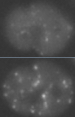
The top image shows a cell before radiation exposure. The bottom image shows the same cell after exposure to 1 Gy of radiation. The bright spots are called radiation induced foci. They indicate the movement of repair proteins toward a site in the cell where DNA damage has occurred.Image Credit: Berkeley LabCostes developed the approach with fellow Berkeley Lab scientists Walter Georgescu, Alma Osserian, Maria Rojec, and Jonathan Tang.
At the heart of their technique are algorithms that lock onto and track individual cells as they move about a cell culture. The algorithms can also follow daughter cells that are created when cells divide. Another component of their approach is the use of human mammary epithelial cells that are modified so that DNA repair proteins, called 53BP1, are fluorescently labeled. This modification, when combined with algorithms that can analyze thousands of cells simultaneously, enables the technique to scan multiple cells and classify areas inside each one where 53BP1 proteins cluster at DNA damage sites. These clusters are called “radiation induced foci.”
The Berkeley Lab scientists have used their cell-tracking and DNA-damage-classification algorithms to analyze human mammary epithelial cells beginning 24 hours before exposure to high and low doses of radiation, and continuing until 24 hours after exposure.
Among their findings is a newly discovered phenomenon: Although DNA damage occurs in random areas throughout a cell, the DNA repair process, as evidenced by the clustering of 53BP1 proteins, is localized in very specific regions of the cell nucleus.
“This could lead to problems,” says Costes. “If the repair process is constrained to specific domains, then there is more of a chance that some of the breaks will meet and get merged together. This would increase the risk of chromosomal rearrangements, such as translocation, where pieces of chromosomes get mixed together, which is considered a precursor to cancer.”
The scientists also found a big difference in how cells respond to DNA damage relative to radiation dose. Some of the differences are well known, such as the fact that high doses of radiation cause cells to stop dividing, whereas low doses don’t arrest cell division. But they also found new processes. At high doses, for example, they discovered that small clusters of 53BP1 proteins merge into larger clusters. This further confirms the risk of chromosomal rearrangement at high doses.
A paper describing their imaging method, with movies of their work, was recently published in the journal PLOS One.
The research was funded by the Department of Energy’s Office of Science.