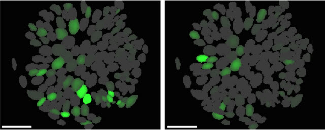
 Each image is of the same exact neurons of a genetically defined group of cells. But some (left) fire while mice search for food; others (right) fire while the mice eat food.Courtesy of Garret Stuber, PhDCHAPEL HILL, NC – Researchers at the UNC School of Medicine have used new deep-brain imaging techniques to link the activity of individual, genetically similar neurons to particular behaviors of mice. Specifically, for the first time ever scientists watched as one neuron was activated when a mouse searched for food while a nearly identical neuron next to it remained inactive; instead, the second neuron only became activated when the mouse began eating.
This work, published in the journal Cell, suggests that manipulating an entire genetically defined subtype of neurons to treat a condition, such as binge-eating, might be too broad of an approach. Drug developers might have to focus on one type of cell within the subset in order to avoid potentially serious side effects.
This study, led by Garret Stuber, PhD, assistant professor of psychiatry, is one of the first published reports using novel technologies that support the NIH BRAIN Initiative to map how individual neurons and neural circuits interact throughout the brain.
 Garret Stuber, PhDMax Englund, UNC School of Medicine“Traditional imaging techniques wouldn’t allow us to record this kind of activity deep inside the brains of freely moving mice,” said Stuber, who is also a member of the UNC Neuroscience Center. “For the first time, we can view specific, genetically defined neurons in the lateral hypothalamus as they light up while the mice search out food, eat, and drink.”
The finding suggests that targeting an entire subpopulation of brain cells to learn about their functions can be somewhat misleading. One type of cell in that subpopulation is somehow predisposed to be involved in one aspect of behavior, while the adjacent neurons are somehow predisposed to be involved in different aspects of behavior.
“This is important to know because if we want to create a drug treatment for obesity, for instance, then you wouldn’t want to affect cells involved in appetite because you might affect cells involved in other aspects of motivated behavior,” said Stuber. “But if we could target only the cells involved in consumption, then maybe we could modulate only those cells without affecting motivation.”
This work, which is part of a larger brain research project in Stuber’s lab, is a good example of how far brain research has come in recent years.
For more than 50 years, scientists have known that basic motivated behaviors, such as eating, drinking, and sleeping, are controlled within the lateral hypothalamus, whether in a rodent, a shark, a human or any other mammal. This part of the brain is very similar in all mammals.
Later studies showed that electrically stimulating the lateral hypothalamus enhanced motivation – the “wanting” of some kind of basic outcome – such as the motivation to eat. Then, researchers found that this brain region is responsible for eating. That is, if there’s food, animals will eat it if this brain region is electrically stimulated. It doesn’t matter if the animals are hungry or not.
But stimulating an entire brain region can’t tell researchers which cell type is truly responsible for which behavior. Until recently, scientists were unable to study these different kinds of cells as they relate to certain kinds of behavior. Stuber decided to use various techniques, including optogenetics and calcium imaging, to study the roles of GABAergic neurons – a large subset of neurons in the lateral hypothalamus.
“By stimulating these cells with light, we found that we could increase feeding and produce reward-related behaviors in mice,” Stuber said. “But, frankly, this finding wasn’t much different than what others found decades ago. We wanted to see whether individual neurons in this one subset of cells were encoding different aspects of behaviors. And this is really challenging. This is why we turned to a specific kind of imaging in live animals.”
Stuber’s team included former UNC Neurobiology Curriculum graduate student Josh Jennings, PhD, and UNC MD/PhD student Randall Ung, who are co-first authors of the Cell paper. They were able to modify only the GABAergic neurons to glow fluorescent when calcium enters neurons – which is what happens during bursts of neuronal activity. Essentially, for the sake of imaging, the fluorescent calcium indicator is a visual proxy for neurons firing.
Because traditional imaging methods do not allow for deep-brain visualization, Stuber’s team turned to a state-of-the-art endoscope imaging system that few other labs have access to. These microscopes, about one inch long, are attached to the brains of mice, which are then able to move and behave normally without restrictions. Stuber’s team was able to study 740 GABAergic neurons in live mice.
Stuber’s team then conducted experiments to analyze the neurons that fired during motivated behaviors, such as searching for food, and the neurons that fired during consummatory behaviors, such as eating and drinking.
When mice searched for food, approximately 22 percent of GABAergic neurons were activated. When the mice consumed food or drink, about 10 percent of the GABAergic neurons fired.
“The key for us is that there was nearly no overlap,” Stuber said. There were cells that only fired during motivated behaviors and cells that only fired during consummatory behaviors.
“When it comes to how these cells function in the brain, we found that there are subpopulations of cells within this larger network of GABAergic neurons,” Stuber said. “And these individual cells are responsible for these highly intertwined activities.
“At some point, we’ll have to be able to target smaller and smaller subpopulations of neurons. The idea is to find genetic markers to delineate these distinct groups of cells. If so, then we could target these groups and learn a lot more about what they do and how they do it.”
This research was funded by the National Institutes of Health, the Klarman Family Foundation, The Foundation for Prader-Willi Research, and the department of psychiatry in the UNC School of Medicine.


