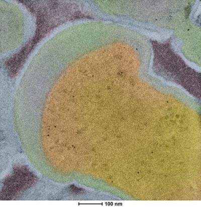
 Ken Kemner is able to track the distribution of bacteria within complex soil aggregates. This colorized image of a thin section of Bacillus subtilis was exposed to conjugated CdSe quantum dots and clearly demonstrates conjugated quantum dots are taken up by the bacteria. This information will be critical for determining the mechanism bacterial cells use to take up these quantum dots.Image credit: by Ken Kemner, Molecular Environmental Science Group at the Argonne National LaboratoryNewswise — Sometimes scientists need space for a little quiet time. At the Environmental Molecular Sciences Laboratory (EMSL), one entire wing of the building was designed to provide just such a place. However, the Quiet Wing wasn’t built to give researchers somewhere to relax. Instead, it was engineered to prevent acoustic noise, vibrations and stray electromagnetic field sources from interfering with the high-resolution capabilities of the imaging instruments housed inside. Under these carefully constructed conditions, seven state-of-the-art microscopes enable scientists to visualize the components of complex samples with unprecedented detail. With information from these atomic-scale images, scientists can advance the understanding of biological and environmental systems.
“The value of these high-resolution imaging instruments is that study systems become a two- or three-dimensional construct,” said Scott Lea, the EMSL Microscopy capability lead. “You can actually see what’s going on, right down to individual atoms.”
In 2012, when EMSL first opened the Quiet Wing, or Q-Wing for short, the microscopes immediately attracted materials and energy storage and conversion types of investigations, Lea noted. Now, EMSL is applying these high-resolution microscopes – two of which also perform compositional analyses – to biological, environmental and aerosol studies that have been considered less traditional uses for such instruments. “These are untapped areas with the potential for a lot of impact,” Lea said.
Quiet Work
Early evidence of that impact came during the Q-Wing’s ramp-up phase. An initial test case for the high-resolution transmission electron microscopes, or TEMs, turned up high-quality images which identified nanoparticulate uraninite being formed by bacteria. The samples came from a decade-old study on uranium remediation led by Ken Kemner, head of the Molecular Environmental Science Group at the Argonne National Laboratory, and EMSL’s Alice Dohnalkova. Although their previous work inferred the presence of isolated ions of reduced uranium from X-ray absorption spectroscopy, at the time there was no technique that could directly image those ions.1 “It was a verification of an idea we put forth almost a decade ago,” said Kemner.
More recently, Kemner and fellow Argonne collaborator Sarah O’Brien use the Q-Wing’s scanning transmission electron microscope, or STEM, to develop approaches to track the distribution of bacteria within complex soil aggregates. The ability to pinpoint the location of bacteria and monitor their activity will improve understanding of soil contaminant transport and fate, the bacterial drivers of carbon dioxide or methane released into the atmosphere, and the effects of microorganisms on plant roots.
However, bacteria ensconced in soil laden with minerals and other opacities can easily dodge detection with traditional X-ray methods. To capture images of bacteria, Kemner is tagging them with fluorescing quantum dots. These dots are nanoparticle-sized pellets of cadmium selenide that are conjugated with amino acids for bacterial uptake.
“With the transmission electron microscope, the Q-Wing is playing a key role in verifying that the quantum dots get into the bacteria via an active uptake mechanism,” Kemner said. Once they nail down that mechanism, Kemner and his colleagues hope to popularize the use of these all-around nano-tracking devices.
Support for this work was provided by the Department of Energy’s Office of Biological and Environmental Research, or DOE BER, specifically, funding was provided by two programs: the Argonne Subsurface Biogeochemical Research Program Scientific Focus Area, which is funded by the DOE BER Subsurface Biogeochemical Research Program; and the Argonne Small Worlds project which is funded by the DOE BER Mesoscale to Molecules Program.
Obtaining atomic-level detail with Q-Wing instruments is also critical to Bradley Tebo, the associate director for the Institute of Environmental Health at Oregon Health and Science University in Portland. With funding from the National Science Foundation’s Chemistry of Life Processes program, Tebo wants to understand the behavior of a unique multicopper oxidase, or MCO, that oxidizes manganese from a soluble Mn+2 state to an insoluble Mn+4.
Unlike other well-characterized enzymes of its ilk, which catalyze single electron transfers, this unusual MCO also requires two accessory proteins to carry out this oxidizing activity in E. coli. Understanding this mechanism may shed light on how manganese conversions occur in the environment, where the availability of the mineral is important for photosynthesis and other key processes, explained Tebo.
“The outstanding scanning transmission electron microscopy capabilities in the Quiet Wing allow us to image single atoms of manganese, and the accessory proteins in the MCO complex,” said Tebo.
As atoms of manganese link with oxygen, this mineral development can be followed on the STEM. With a time series of images taken during the reaction, Tebo hopes to determine whether the minerals immediately form a well-developed structure, or create an amorphous shape that later organizes into orderly sheets. He can also examine the accessory proteins to see how they fold and where the protein subunits are bound to the MCO. Ultimately, he wants to discover whether the protein complex itself serves as a template for mineral formation, or whether some other control exists.
Once the mineralization process is understood, it might be modified to perform tasks such as targeted metal sequestration or – with a slightly altered redox potential – perhaps create a renewable catalyst that can directly deoxidize water to form hydrogen.
Such fine structural detail is equally important to see what happens to the sponge-like properties of clay after it’s coated with natural organic material and exposed to water. Interactions at these dirty surfaces play key roles in remediation methods using porous clay to soak up environmental contaminants or in shale-based sequestration techniques that use clay pores in the shale rock as carbon storage chambers, explained Geoffrey Bowers, an associate professor of chemistry at Alfred University in New York.
After many years of exploring the properties of hectorite (a clay with minimal iron content) with EMSL’s experts in nuclear magnetic resonance, or NMR, and X-ray diffraction, Bowers made “a concerted shift” to study the interfaces between clay and natural organic matter, or NOM. This research is supported by funds from the DOE’s Office of Basic Energy Sciences, Geosciences program.
“We have reasonable ideas about how pristine clay affects the transport of ions in the near surface soils and we know organic matter is good at picking up heavy metals like iron and arsenic,” said Bowers. “But it’s important to know what happens when you put them together because in surface soil you’re almost never going to find clay that’s not in intimate contact with NOM.”
With his longtime collaborator, R. James Kirkpatrick, dean of the College of Natural Science at Michigan State University, Bowers turned to the Q-Wing’s helium ion microscope, or HIM, to get a more holistic view of this complex system. The HIM is similar in concept to a scanning electron microscope, but has higher spatial resolution, greater depth of field and increased sensitivity to small changes at the clay surface. With this instrument the researchers could study natural organic matter-clay composites at scales ranging from tens of nanometers up to one micron in size.
There’s a long list of questions Bowers wants to answer with the HIM. For example: Do the samples have large amounts of organic matter preventing water or ions from accessing the clay fraction? Do the clays interact with NOM? What does the pore network look like before and after NOM is introduced? Are there chunks of hydrophobic organic matter that won’t let water in, or do super-porous organic matter flocculent particles soak up more water than we expect?
“Those are questions we can’t answer with molecular-scale techniques we typically use such as molecular dynamics modeling or NMR,” said Bowers. “But we need the answers to properly interpret our models and NMR results. That’s where the Q-Wing comes into play.”
So far, Bowers has seen subtle surface changes that couldn’t be captured with electron microscopy alone. For example, the basal surfaces of clay appear flat under the HIM lens. However, the composite material surfaces show “little hills and hummocks” that suggest there’s a coating on the clay, said Bowers. Going forward, he’ll study how altering the chemical conditions of clay-NOM composite suspensions influence that structure.
Another surprise from the Q-Wing studies was that some composite samples, under certain conditions, form “taffy-like strands” of NOM between the clay particles. These strands look like a “string of pearls,” said Bowers, with the size of each pearl in the string correlating with other research results which suggest small 10-20 nm globules may be the state of NOM in solution. With further study, the types of structures formed in clay-NOM composites could be manipulated to improve targeted ion absorption.
Bowers and his colleagues are also exploring subsurface samples with Q-Wing microscopes. “We want to know what might happen if supercritical carbon dioxide is pumped into shale to sequester it,” he said. “Will the ions in clay help form more stable mineralized carbon phases?”
There’s no endpoint to his questions about these materials and how they affect transport, with or without supercritical CO2 around, said Bowers. “I think we’ve opened the lid on a treasure chest, and the question is how far we are going to reach in there and pull out gold for the scientific community as a whole.”
Breaking the Silence
Spreading the word about these Q-Wing capabilities is done primarily through meetings, said Lea. “Either we’re presenting our own work, or talking with investigators to understand what they’re trying to do and see how we might be able to help them,” he said.
Soon, scientists using the Q-Wing will have even better options available to study their research samples.
Efforts by Nigel Browning, at Pacific Northwest National Laboratory, and James Evans in EMSL are underway to develop a second-generation dynamic TEM that allows faster time resolution (in the nanosecond realm) as well as high spatial resolution. “We’re trying to go three to six orders of magnitude faster,” said Lea. “If we can do that, then we’ll be able to see things like protein conformational dynamics such as helix or domain motions, aerosol growth and nucleation. There’s no way to get this information now using electron-based approaches.”
EMSL experts are also planning to add high-sensitivity, direct electron detector devices, or DEDs for short, on some of the Q-Wing instruments. Replacing the charge-coupled devices currently on the TEMs with these new devices will give microscopists high-resolution images with a smaller dose of electrons. The DEDs will be particularly advantageous for aerosol or biological samples that are more sensitive to beam damage caused by exposure to large doses of electrons. The liquid helium cryogenic transmission electron microscope is the first instrument slated for getting the new device, Lea said.
Another technique championed by the Chemical Imaging Initiative at PNNL that would give researchers more imaging information with fewer electrons is a mathematical method called compression sensing. This method provides similar information, but with less electron dose, by reducing the noise in the images.
“These combined efforts should give us the ultimate in low-dose imaging, which is very important going forward for biochemical, environmental and aerosol research,” said Lea. Still, he’s eager to fill one empty instrument berth in the Q-Wing with the next advanced instrumentation.
Reference:
1. Dohnalkova A, Marshall M, Kemner K, Kennedy D, Genc A and Fredrickson J. 2014. “Bacterially-Produced Uraninite – Ultrastructural Characterization by High Resolution Imaging and Analyses.” Goldschmidt conference in Sacramento, Calif.