Cell Biology

The evolution of organoids as next-generation research and screening models
| 5 min read

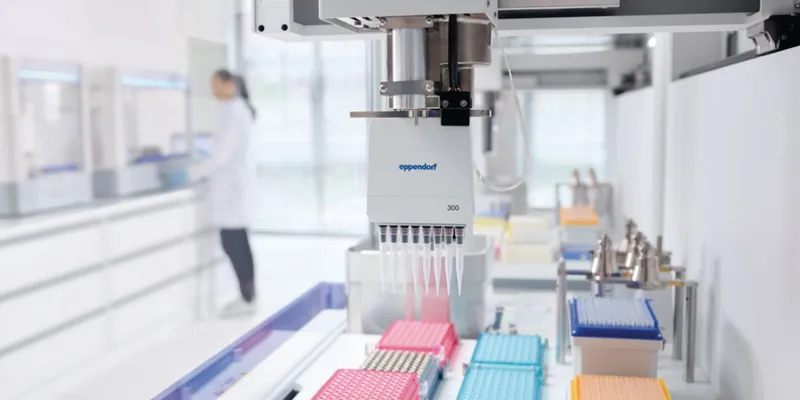
A fully automated flow cytometry sample preparation system
Updated | 2 min read

New multimodal approach combines XCT and XRF imaging to reveal detailed cell structures and processes
| 5 min read

Poking holes through membranes with atomic accuracy to build pocket-sized ‘molecule detectives’
| 3 min read

Research collaboration achieves scientific breakthrough in understanding cell division
| 2 min read

Learn how automated whole blood sample preparation improves immunophenotyping accuracy and efficiency
| 1 min read

Researchers unlock crucial molecular secrets of aging in cells, potentially paving the way to improve quality of life
| 2 min read

Findings alter previous scientific views and may be relevant to skin cancer development
| 3 min read

Researchers have developed a method to precisely target cells for inflammatory cell death using light
| 2 min read

Research sheds new light on marine organisms' ability to adapt and survive in the ocean
| 5 min read