Cell Biology

Unveiling the proteins behind cellular organelle communication
| 2 min read

Discovery challenges textbook notions, provides insights on eukaryotic evolution and global agricultural potential
| 4 min read

Researchers propose that the existence of parallel lineages influences the folding of the cerebral cortex
| 3 min read

Unlocking successful growth: incubator maintenance and care
Updated | 1 min read

High-throughput Nanowell-based Image Verified Cloning technology sets the stage for accelerated generation of clonal pharmaceutical production cell lines
Updated | 1 min read

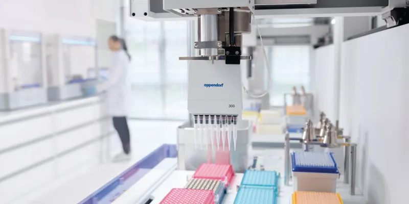
Manual and automated liquid handling systems are not immune to error but this can be rectified with effective protocols, regular maintenance, and proper training
Updated | 1 min read

Researchers pioneer blastoid models to study earliest stages of embryonic development, aiding future therapies and treatments
| 3 min read

Stable and non-toxic fluorescent dye allows for live visualization of membrane fluidity during cellular processes
| 3 min read

Researchers have discovered a mechanism steering the evolution of multicellular life
| 2 min read

Researchers shed light on archaea, a single-cell microorganism, and the molecular mechanisms behind cell shape formation
| 4 min read