
Fluorescence

As microscopy reporting requirements tighten, imaging cores face growing pressure to demonstrate reproducibility. These five questions can help identify potential gaps in quality control, performance monitoring, and documentation workflows
| 2 min read

| 1 min read

Updated | 5 min read
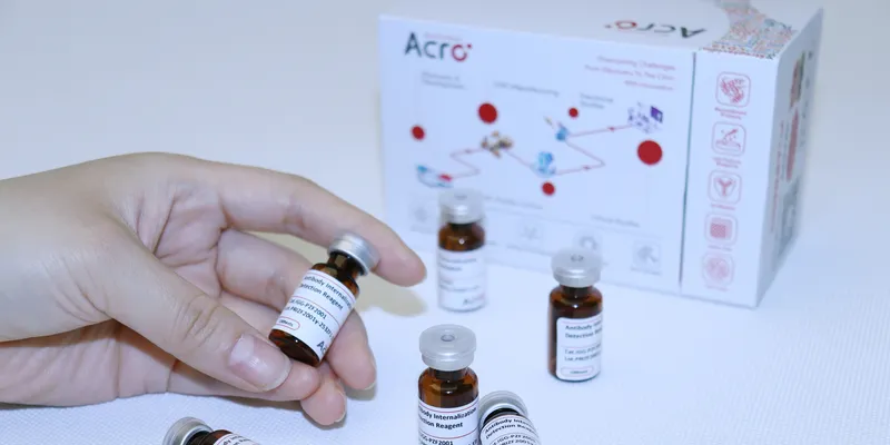
This pH-sensitive fluorescent Fc-label enables real-time tracking of antibody–drug conjugate (ADC) uptake in live cells with strong signal and low background, ideal for studying internalization by flow cytometry or fluorescence imaging
| 1 min read

Learn how to properly and safely use a fluorescence microscope, from setup to image capture, with our comprehensive step-by-step guide
Updated | 4 min read

Versatile readers and automation technology support a wide range of applications
| 1 min read

The InverTau™ platform simplifies fluorescence lifetime imaging measurements
| 2 min read

Researchers use satellite imagery to predict crop yields by measuring solar-induced chlorophyll fluorescence
| 3 min read

Newly developed SR fluorescence microscopy technique enables more accurate mapping at an unparalleled scale
| 2 min read

Introducing the upgraded SPECTRO XEPOS, with excellent performance at much lower cost
| 2 min read