Fluorescence

The LIFA vTAU is particularly helpful for live cell applications
| 1 min read

Understanding the mechanics of neural function can help treat neurodegenerative diseases
| 5+ min read

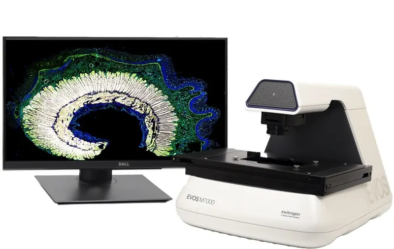
Rapid advancements mark an expanding frontier in life sciences research accessible to all
| 5+ min read

A handbook for everyone, from beginners to experts
| 1 min read

Recent technological leaps improve sustainability, uniformity, and control in imaging at a reduced cost
| 2 min read

The Gel-Bright™ Laser Diode Gel Illuminator is more sensitive than LED illuminators for red dyes, without UV hazards
| 2 min read

The technique works for bacterial cell division and for mitochondrial division
| 2 min read

Secondary Antibodies for high-resolution imaging
| 1 min read

Bringing the western blot into the 21st century
| 1 min read