Trace evidence in forensic science covers vast territory, including microscopic signs of gunshots and even strands from a wig. To analyze such different types of samples, scientists must use a variety of microscopes.
“Trace evidence is material that can be transferred during the commission of a crime,” says Vince Vaccarelli, product manager for forensics and education at Leica Microsystems (Buffalo Grove, IL). “Examples are hair, fiber, paint, body fluids, chemicals, or any other particles left behind during a crime.” He adds, “That means you must be prepared to use a variety of contrast techniques, lighting, and magnifications to best suit the evidence you are studying.”
When it comes to applying microscopy to forensics, Donna Guarrera, scanning electron microscope product manager at JEOL (Peabody, MA) says, “You start with a stereo- or light microscope, and then you advance to electron microscopy if you need to look on the micron, submicron, or even nanometer level—depending on the microscope.” In addition to providing higher levels of magnification, electron microscopy (EM) delivers more depth of field—sometimes 2-3 orders of magnitude better than light microscopy.
Sign up to receive the Forensic Science Tools and Techniques Newsletter
In many cases, researchers combine other technologies with microscopy. “Nearly every analytical tool used in a trace-evidence lab has been adapted as an accessory to the microscope,” says Christopher Palenik, vice president and senior research microscopist at Microtrace (Elgin, IL). As examples, he mentions infrared and Raman spectroscopy. “In our lab, light microscopy—mainly the stereomicroscope and polarized light microscope—represents a fundamental starting line for all samples,” he says. “The results of these initial microscopical surveys direct the use of other, more specialized microanalytical tools.”
Preparing for imaging
When asked how to prepare a forensic sample for microscopy, Palenik says, “Carefully.” He adds that forensic samples are rarely pure, and most analytical methods work best with pure or nearly pure samples.
So, samples must be prepared as needed. Some samples—like fibers or wood—can be isolated under a stereomicroscope. “Microscale separations of compounds through solubility or chromatography represents a way to purify trace residues or liquids,” Palenik says. “The use of specialty preparation methods—such as microtomy, polishing, and ion milling—are becoming increasingly important as trace evidence looks to smaller scales and more precise results.”
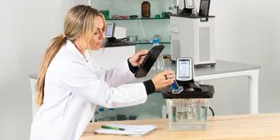
A sample that will be analyzed with transmitted light microscopy is prepared on a glass slide using a mounting medium. “Mounting media examples are immersion oils, liquids of different refractive index, or optical cement,” Vaccarelli explains. Other forms of microscopy require different methods of sample preparation.
EM explorations
Beyond looking at samples, EM can analyze their chemistry. “When you hit a sample with a high-energy beam of electrons, many signals are generated, and you can get elemental information on a sample,” says Guarrera.
Take gunshot residue as an example. Here, forensic scientists rely almost entirely on EM. The particles are 1-10 microns in size (a human hair is 100 microns in diameter). Using scanning EM (SEM) with a backscatter detector, a scientist can scan a sample to reveal high-density materials. The high-density spots can be analyzed with energy dispersive spectroscopy to look for signs of the elements antimony, barium, and lead, which come from the primer of a fired bullet.
As David Edwards—senior applications specialist at JEOL, who is also on the Organization of Scientific Area Committees’ gunshot residue subcommittee—explains this process: “Primer residue vaporizes and becomes molten, and this will coalesce into a microscopic sphere that we look for.”
This area, though, shows the need for methods to evolve. For example, Europe is removing lead even from bullet primer. So, the search signature must be changed accordingly.
Many applications of EM in forensics go beyond gunshots. For example, paint transferred between vehicles in a crash can be analyzed to unimaginable depths. “With SEM, you can look at how many layers are in the transferred paint and get some idea of what the pigments are,” says Guarrera. In fact, vehicle manufacturers provide paint details for forensic databases. This information includes the specific layer structure, such as the thickness and the number of layers for a specific manufacturer and model. These features can also be explored with other forms of imaging.
As much as the applications expand, so does EM technology. Most of all, it is easier to use. “A forensic scientist or biologist doesn’t have to be an expert in EM anymore,” Guarrera notes. “We have a three-day training course, and that’s even enough for people with no exposure to this technology.” In fact, modern EM is easy enough to use that JEOL installed one in a high school.
As Edwards explains, “Instead of needing a dedicated operator, EM is now a tool in the toolbox.” Some platforms operate like a tablet, and even sample preparation is simplified considerably versus a few decades ago. When using low-vacuum mode, for instance, nonconductive samples no longer need a conductive coating.
Beyond a hair of a doubt
Any kind of sample at a crime scene could be used as evidence. Hair is often used, and it can even come from a wig. At Sam Houston State University (Huntsville, TX), Patrick Buzzini, associate professor in forensic science, used light microscopy to examine wig fibers.
In particular, Buzzini and his colleagues examined the crosssectional shape of the fibers. “We hypothesized that this feature is extremely variable between samples from different origins,” he explains. Moreover, he wanted an analytical approach to compare unknown wig fibers with ones from a known source, such as a wig from a crime scene.
Buzzini’s team categorized wig-fiber samples based on 10 types of cross-sectional shapes. “We then correlated the cross-sectional shapes with the chemical composition obtained using infrared spectroscopy,” he says. In that way, Buzzini showed that the wig fibers could be compared based on shape and composition.
From wig fibers to micrometer residue from a gunshot, forensic scientists apply microscopy in many ways. The method can provide images, but it can also go deeper, such as enabling collection of chemical information. Any results from microscopy of trace elements could make the difference in a case’s outcome.


