In the modern scientific and industrial landscape, the ability to precisely and thoroughly understand materials is not merely a supplementary activity; it is a foundational requirement for innovation, quality assurance, and failure analysis. Materials characterization stands as the cornerstone of this understanding, providing a comprehensive assessment of a material’s composition, structure, and properties. It is a critical discipline that underpins nearly every field of study, from the development of novel pharmaceuticals and high-performance composites to the validation of semiconductor technologies and the analysis of environmental samples. The insights gained from materials characterization directly influence material performance, longevity, and safety, making it an indispensable part of any laboratory workflow.
For laboratory professionals, a mastery of advanced material analysis techniques is essential for validating hypotheses, ensuring product consistency, and adhering to strict regulatory standards. The transition from traditional methods to more sophisticated, high-resolution techniques has enabled unprecedented insights into materials at the atomic and molecular levels. This article explores the core principles and advanced methodologies that define modern materials characterization, offering a guide to the techniques that are shaping the future of scientific discovery and industrial application. It provides an authoritative overview of how these methods are integrated to provide a holistic understanding of materials, reinforcing their professional significance in contemporary laboratory settings.
Mastering Foundational Principles for Modern Materials Characterization
The essence of materials characterization lies in its ability to link a material's intrinsic properties to its behavior and performance. This is achieved by systematically investigating several key attributes: chemical composition, atomic and molecular structure, microstructure, and surface morphology. A single analytical technique rarely provides all the necessary information; instead, a multi-modal approach is often required, combining various material analysis techniques to build a complete picture.
The selection of appropriate analytical methods depends on the specific question being asked. For instance, determining the elemental composition of a sample necessitates techniques like Energy Dispersive X-ray Spectroscopy (EDS) or X-ray Photoelectron Spectroscopy (XPS), while understanding crystalline structure requires X-ray Diffraction (XRD). Characterizing surface roughness or morphology, on the other hand, calls for advanced forms of microscopy. This integration of complementary data streams is what elevates materials characterization from a series of individual tests to a cohesive and powerful analytical process.
An effective materials characterization workflow begins with careful sample preparation, as the quality of the sample can dramatically affect the results. This is followed by the application of one or more primary techniques. The data from these initial analyses often informs the need for secondary, more specialized techniques to address specific anomalies or to obtain higher-resolution information. The final step involves a rigorous interpretation of all collected data, cross-referencing findings from different techniques to arrive at a conclusive and validated characterization of the material. This structured, logical process is crucial for ensuring the reproducibility and integrity of scientific outcomes in a professional laboratory environment.
Advanced Microscopy and XRD for Material Structure Analysis
The ability to visualize a material's physical structure is a fundamental aspect of materials characterization. This is where advanced microscopy and diffraction techniques provide invaluable insights into a material's morphology, grain size, and crystalline orientation.
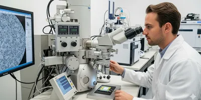
- Scanning Electron Microscopy (SEM): SEM provides high-magnification images of a sample's surface topography. A focused beam of electrons is scanned across the surface, and the resulting secondary and backscattered electrons are detected to form an image. This technique is non-destructive and highly effective for studying a wide range of materials, including metals, polymers, and biological specimens. The high depth of field in SEM images provides a three-dimensional perspective, revealing surface features with remarkable clarity.
- Transmission Electron Microscopy (TEM): For a more detailed look into a material's internal structure, TEM is a powerful tool. In TEM, a beam of electrons is transmitted through an ultra-thin sample. The interactions between the electrons and the sample’s atoms provide information about the material’s crystal structure, defects, and morphology at the nanoscale. TEM can achieve resolutions far superior to SEM, enabling the visualization of individual atoms within a crystal lattice, which is crucial for advanced materials research.
- Atomic Force Microscopy (AFM): Unlike electron microscopy, AFM does not use an electron beam. Instead, it employs a cantilever with a sharp tip to physically "feel" the surface of a sample. By measuring the forces between the tip and the sample, AFM can generate a three-dimensional map of the surface topography with angstrom-level resolution. This makes it an ideal technique for analyzing delicate biological samples and soft materials that could be damaged by an electron beam.
- X-ray Diffraction (XRD): For crystalline materials, XRD is the gold standard for structural analysis. When X-rays interact with a crystalline solid, they are diffracted by the planes of atoms in the crystal lattice. According to Bragg’s Law, specific angles of incidence produce constructive interference, resulting in sharp peaks in the diffraction pattern. Each crystalline material produces a unique diffraction pattern, which acts as a "fingerprint" for its atomic structure. XRD can be used for phase identification, determining lattice parameters, quantifying crystallinity, and analyzing crystallite size. It is a fundamental technique for quality control in the pharmaceutical, chemical, and mineral industries.
The synergy between these techniques is highly effective. For example, SEM might be used to observe the overall morphology of a material, while XRD provides data on its crystalline phases. Then, TEM could be used to probe the structure of individual crystallites at high resolution, and AFM could be employed to study the surface roughness of a specific area. This layered approach ensures a thorough and validated materials characterization.
Spectroscopy: Probing Material Composition and Purity
Spectroscopic techniques are essential material analysis techniques for understanding the chemical composition and bonding of a material. They involve measuring the interaction between electromagnetic radiation and matter. Each technique is sensitive to a different aspect of a material’s chemical identity.
- Fourier-Transform Infrared (FTIR) Spectroscopy: FTIR is a rapid and versatile technique for identifying functional groups and chemical bonds within organic and inorganic compounds. It works by passing infrared light through a sample and measuring how much light is absorbed at different wavelengths. The resulting spectrum, a plot of absorbance versus wavenumber, provides a unique fingerprint for the material. FTIR is widely used in polymer science, pharmaceutical quality control, and forensic analysis.
- Raman Spectroscopy: Similar to FTIR, Raman spectroscopy provides information about molecular vibrations and rotational modes. However, it relies on inelastic scattering of light, where the scattered photons have a different energy than the incident photons. Raman spectroscopy is particularly useful for analyzing carbon-based materials like graphene and carbon nanotubes, as well as minerals and certain biological samples. It can also be performed through transparent containers, making it ideal for non-invasive analysis.
- X-ray Photoelectron Spectroscopy (XPS): XPS is a powerful surface-sensitive technique that provides both elemental composition and chemical state information. The sample is irradiated with X-rays, causing the ejection of core-level electrons. By measuring the kinetic energy of these electrons, it is possible to determine the elemental composition of the top few nanometers of the surface. Furthermore, shifts in the binding energies of the electrons reveal information about the chemical environment of each element, such as its oxidation state. XPS is an essential tool for studying thin films, catalysts, and surface contaminants.
- Energy Dispersive X-ray Spectroscopy (EDS/EDX): Often integrated into electron microscopes like SEM and TEM, EDS is used for elemental analysis. The electron beam excites atoms in the sample, causing them to emit characteristic X-rays. The energy and intensity of these X-rays are unique to each element, allowing for a qualitative and quantitative analysis of the sample’s elemental composition. EDS is a quick and straightforward method for identifying the constituent elements of a material at the microscopic level. The integration of EDS with microscopy instruments provides a powerful capability to correlate morphology with elemental composition in real-time.
Synergizing Material Analysis Techniques for Comprehensive Results
Modern materials characterization is defined by its integrated approach. Seldom does a single technique provide a comprehensive answer; rather, the combination of complementary methods allows for a holistic and validated understanding of a material. This synergy is particularly important in fields where complex interactions between composition, structure, and morphology dictate a material’s performance.
Consider the analysis of a a novel composite material designed for aerospace applications. A possible workflow would be as follows:
Initial Assessment with Microscopy: An SEM would first be used to examine the composite’s surface and cross-section, revealing the distribution of reinforcing fibers within the polymer matrix. This provides initial insights into the overall integrity and uniformity of the material.
Structural and Crystalline Analysis: An XRD experiment would then be conducted on the composite. This would provide information on the crystallinity of both the polymer matrix and the reinforcing fibers, as well as any crystalline phases that might have formed at the interface between the two components.
Chemical Composition and Bonding: FTIR and Raman spectroscopy could be used in parallel to identify the chemical composition of the polymer matrix and the fibers. FTIR would be sensitive to the overall bulk chemistry, while Raman could provide more specific information about the molecular structure of the carbon fibers.
Surface Chemistry and Contaminants: A final, high-resolution analysis could involve XPS. By analyzing the surface of the composite, XPS could identify any surface treatments applied to the fibers and detect any contaminants that could compromise the bond between the fiber and the matrix.
This integrated approach yields a far richer and more reliable dataset than any single technique could provide. It allows researchers to correlate a material’s performance metrics (e.g., tensile strength or thermal stability) with specific structural or chemical features, providing a direct link between processing, structure, and performance. This holistic view is essential for both basic research and for diagnosing failures in industrial applications.
Elevating Laboratory Practice with Advanced Materials Characterization
The field of materials characterization continues to evolve, driven by advancements in instrumentation, data processing, and analytical methodologies. For laboratory professionals, keeping pace with these developments is not merely an academic exercise; it is a professional imperative. A deep understanding of the fundamental principles of microscopy, spectroscopy, and XRD, as well as the ability to strategically integrate these material analysis techniques, is what separates routine analysis from true scientific inquiry.
The professional significance of this discipline extends across every stage of the product lifecycle. From the initial research and development of a new material to the quality control of a final product and the post-mortem analysis of a failed component, materials characterization provides the quantitative data necessary for informed decision-making. By mastering these advanced techniques, laboratory professionals can contribute to a deeper understanding of materials, drive innovation, and ensure the safety and reliability of the products that shape modern society. Continued education and a commitment to adopting new technologies are paramount for those who wish to remain at the forefront of this critical and dynamic field.
FAQ
How does spectroscopy differ from microscopy in materials characterization?
Spectroscopy and microscopy are distinct but often complementary material analysis techniques. Spectroscopy primarily focuses on the interaction of matter with electromagnetic radiation to provide information about a material’s chemical composition, elemental makeup, and molecular bonding. Techniques like FTIR and XPS analyze the energy absorption or emission patterns to identify specific chemical functional groups or elements. In contrast, microscopy is a direct visualization tool that provides high-resolution images of a material's physical structure, morphology, and topography. Electron microscopes (SEM, TEM) and atomic force microscopes (AFM) are used to observe features such as particle size, grain boundaries, and surface defects. While microscopy answers the question "what does it look like?", spectroscopy answers the question "what is it made of?". The most powerful materials characterization workflows integrate both to provide a holistic understanding.
What is the significance of X-ray Diffraction (XRD) in the broader context of material analysis techniques?
XRD holds a unique and crucial position among material analysis techniques due to its specific focus on crystalline structure. While many other methods provide information on composition or morphology, XRD is the definitive tool for identifying the crystalline phases present in a sample, determining their relative quantities, and measuring parameters such as lattice dimensions and crystallinity. This is invaluable for quality control in industries where crystal structure is directly linked to performance, such as in pharmaceuticals, where polymorphism can affect drug efficacy, or in metallurgy, where specific crystal structures are required for certain mechanical properties. XRD’s ability to provide a "fingerprint" of a crystalline material makes it an indispensable tool for both unknown sample identification and for the verification of material authenticity and purity.
Why is an integrated approach to materials characterization considered a best practice?
An integrated approach to materials characterization is considered a best practice because no single technique can provide a complete picture of a material's properties. A material's performance is a function of its composition, structure, and morphology, and each of these attributes requires different material analysis techniques to be fully understood. A simple example illustrates this: a change in a material's thermal stability might be due to a subtle change in its chemical composition (detectable by spectroscopy), a change in its crystalline structure (detectable by XRD), or a change in its microstructure (detectable by microscopy). By using a combination of methods, laboratories can identify the root cause of a specific material behavior or property, leading to more accurate conclusions, effective problem-solving, and more robust research and development outcomes. This holistic perspective is essential for both fundamental research and industrial failure analysis.
How can laboratory professionals ensure the accuracy and reliability of their materials characterization data?
Ensuring the accuracy and reliability of materials characterization data is paramount for maintaining scientific integrity and meeting industry standards. This begins with meticulous sample preparation, as a poorly prepared sample can lead to misleading results regardless of the sophistication of the instrumentation. It is also critical to perform regular instrument calibration and maintenance to ensure that all equipment is operating within its specified performance parameters. Data acquisition protocols should be standardized and consistently followed to minimize operator-induced variability. Finally, data analysis and interpretation should involve a rigorous cross-referencing of results from multiple material analysis techniques. For instance, if spectroscopy suggests a certain chemical bond is present, that finding should be corroborated by the structural information provided by XRD or high-resolution microscopy. This multi-faceted validation process is key to producing reliable and defensible characterization data.


