Analytical Instruments

The new portfolio features both compound and stereozoom microscopes for all your needs
| 1 min read

Innovation gives researchers inside view of how batteries work
| 3 min read

The DynaGreen™ Magnetic Beads can help labs adopt more sustainable practices
| 1 min read

Effects of ionizable and non-ionizable excipients on lyophilized RNA formulations using FTIR-ATR technology
| 1 min read


The LCMS-9050 system builds upon the innovative technologies developed for the widely used Shimadzu LCMS series
| 1 min read

The new tool, Magnify, is a potent and accessible method that can provide new insights into a variety of fields
| 4 min read

The open-source software is the first metabolite identification tool that can distinguish between stereochemical variants
| 2 min read

Deep learning can help in characterizing materials by analyzing peak changes in x-ray diffractions
| 2 min read

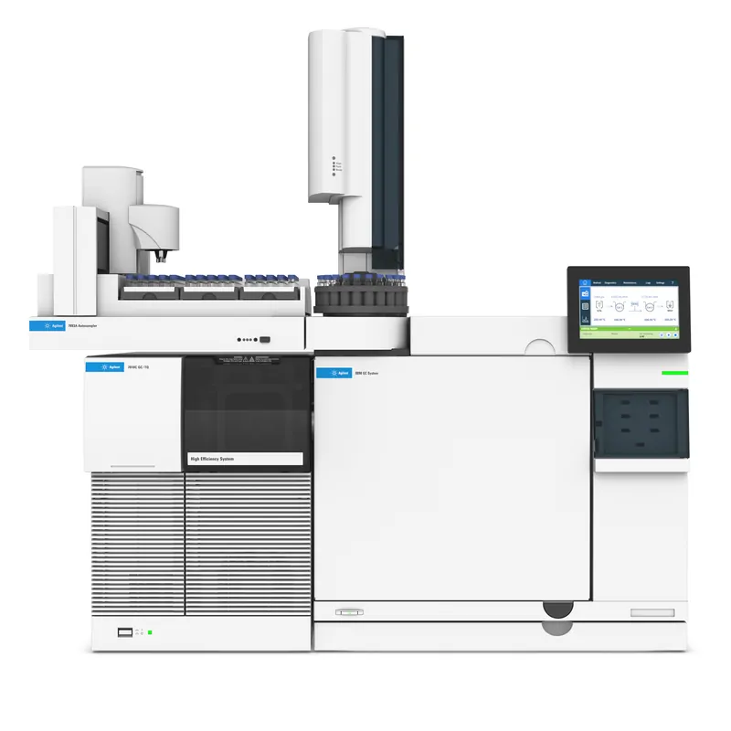
A new hydrogen carrier gas electron ionization (EI) source designed to improve chromatographic efficiencies for your gas chromatography/mass spectrometry (GC/MS) workflow
| 4 min read

The Agilent 7000E triple quadrupole GC/MS provides unequivocal intelligence and robustness to deliver the answers you seek.
| 2 min read
The Agilent 7010C triple quadrupole GC/MS is the most sensitive instrument of the Agilent GC/TQ portfolio that is powered by ultimate mass spec intelligence.
| 1 min read


