Microscopy
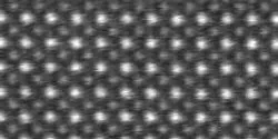
In July 1978, Peter Buseck of Arizona State University, together with two postdoctoral researchers (also then at ASU), published a paper on a new technique for high-resolution imaging of crystal structures using transmission electron microscopes. Recently, the scientific journal Nature has hailed that paper as a milestone in the science of crystallography. At the same time, Nature also cited three other milestone crystallography papers.

A research group led by Professor Hiroyuki Noji, Department of Applied Chemistry, Graduate School of Engineering, University of Tokyo, successfully observed and touched the rotational motion of a 1-nm synthetic molecular machine through the application of a single-molecule capturing and manipulation technique using optical microscopy and a bead probe (single-molecule motion capturing), which allows visualization of molecular mechanical motion.

Researchers from Carnegie Mellon University have developed a novel method for creating self-assembled protein/polymer nanostructures that are reminiscent of fibers found in living cells. The work offers a promising new way to fabricate materials for drug delivery and tissue engineering applications. The findings were published in the July 28 issue of Angewandte Chemie International Edition.
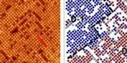
A novel combination of microscopy and data processing has given researchers at the Department of Energy’s Oak Ridge National Laboratory an unprecedented look at the surface of a material known for its unusual physical and electrochemical properties.

Ferroelectric materials–substances in which there is a slight and reversible shift of positive and negative charges–have surfaces that are coated with electrical charges like roads covered in snow. Accumulations can obscure lane markings, making everyone unsure which direction traffic ought to flow; in the case of ferroelectrics, these accumulations are other charges that “screen” the true polarization of different regions of the material.

Dr. Zhaozheng Yin, an assistant professor of computer science at Missouri University of Science and Technology, recently received the National Science Foundation’s most prestigious award for young faculty members for his research into developing algorithms and systems for processing microscopy images of biological specimens.

