Microscopy

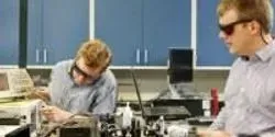
Thermal imaging, microscopy and ultra-trace sensing could take a quantum leap with a technique developed by researchers at the Department of Energy’s Oak Ridge National Laboratory.

UT Southwestern Medical Center’s Texas Institute for Brain Injury and Repair (TIBIR) has acquired a pair of TissueCyte 1000 microscopes, the latest generation in serial two-photon laser imaging, as a centerpiece of its new Whole Brain Microscopy Facility. The TissueCyte microscopes are the only ones of their kind in Texas, and two of just a handful in existence around the world.

Scientists at Albert Einstein College of Medicine of Yeshiva University and their international collaborators have developed a novel fluorescence microscopy technique that for the first time shows where and when proteins are produced. The technique allows researchers to directly observe individual messenger RNA molecules (mRNAs) as they are translated into proteins in living cells. The technique, carried out in living human cells and fruit flies, should help reveal how irregularities in protein synthesis contribute to developmental abnormalities and human disease processes including those involved in Alzheimer’s disease and other memory-related disorders. The research will be published the March 20 edition of Science.

Vanderbilt University researchers have achieved the first “image fusion” of mass spectrometry and microscopy — a technical tour de force that could, among other things, dramatically improve the diagnosis and treatment of cancer.

Advances in computer hardware and software, data storage and processing, optics, systems and instrumentation, labeling agents, and reagents have all contributed to the current surge in imaging in the life sciences.

Delivering the capability to image nanostructures and chemical reactions down to nanometer resolution requires a new class of x-ray microscope that can perform precision microscopy experiments using ultra-bright x-rays from the National Synchrotron Light Source II (NSLS-II) at Brookhaven National Laboratory. This groundbreaking instrument, designed to deliver a suite of unprecedented x-ray imaging capabilities for the Hard X-ray Nanoprobe (HXN) beamline, brings researchers one step closer to the ultimate goal of nanometer resolution at NSLS-II, a U.S. Department of Energy Office of Science User Facility.

Dr. Anne Carpenter leads the Imaging Platform at the Broad Institute of Harvard and MIT—a team of biologists and computer scientists who develop image analysis and data mining methods and software that are freely available to the public through the open-source CellProfiler project.
Dr. Arvind Rao has been an assistant professor in the Department of Bioinformatics and Computational Biology at the University of Texas MD Anderson Cancer Center since 2011.


