Analytical Instruments

Inductively coupled plasma optical emission spectrometry (ICP-OES) is used for elemental analysis of everything from soil and sludge to water and wastewater, plus various industrial process materials. In evaluating ICP-OES instruments, environmental contract laboratories may prioritize sensitivity and speed. Industrial research laboratories may emphasize stability and analytical precision. However, both agree on the importance of controlling costs.

Dr. Anne Carpenter leads the Imaging Platform at the Broad Institute of Harvard and MIT—a team of biologists and computer scientists who develop image analysis and data mining methods and software that are freely available to the public through the open-source CellProfiler project.
Dr. Arvind Rao has been an assistant professor in the Department of Bioinformatics and Computational Biology at the University of Texas MD Anderson Cancer Center since 2011.

Opening new doors for biomedical and neuroscience research, Elizabeth Hillman, associate professor of biomedical engineering at Columbia Engineering and of radiology at Columbia University Medical Center (CUMC), has developed a new microscope that can image living things in 3D at very high speeds. In doing so, she has overcome some of the major hurdles faced by existing technologies, delivering 10 to 100 times faster 3D imaging speeds than laser scanning confocal, two-photon, and light-sheet microscopy.

JEOL USA and the University of California's Irvine Materials Research Institute (IMRI) have entered into a strategic partnership to create a premier electron microscopy and materials science research facility. The IMRI will serve as an interdisciplinary nexus for the study and development of new materials, enabling advances in solar cell, battery, semiconductor, biological science, and medical technologies.

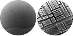
Rust never sleeps. Whether a reference to the 1979 Neil Young album or a product designed to protect metal surfaces, the phrase invokes the idea that corrosion from oxidation—the more general chemical name for rust and other reactions of metal with oxygen—is an inevitable, persistent process. But a new study performed at the Center for Functional Nanomaterials (CFN) at the U.S. Department of Energy's (DOE) Brookhaven National Laboratory reveals that certain features of metal surfaces can stop the process of oxidation in its tracks.

Ames Laboratory scientists use genetic markers to discover the rhizosphere.

Scientists at the Department of Energy’s Oak Ridge National Laboratory have used advanced microscopy to carve out nanoscale designs on the surface of a new class of ionic polymer materials for the first time. The study provides new evidence that atomic force microscopy, or AFM, could be used to precisely fabricate materials needed for increasingly smaller devices.


