Imaging

UT Southwestern Medical Center’s Texas Institute for Brain Injury and Repair (TIBIR) has acquired a pair of TissueCyte 1000 microscopes, the latest generation in serial two-photon laser imaging, as a centerpiece of its new Whole Brain Microscopy Facility. The TissueCyte microscopes are the only ones of their kind in Texas, and two of just a handful in existence around the world.

A research team led by Shree K. Nayar, T.C. Chang Professor of Computer Science at Columbia Engineering, has invented a prototype video camera that is the first to be fully self-powered—it can produce an image each second, indefinitely, of a well-lit indoor scene. They designed a pixel that can not only measure incident light but also convert the incident light into electric power. The team is presenting its work at the International Conference on Computational Photography at Rice University in Houston, April 24 to 26.

Bestselling author Sam Kean takes us through the nearly 2,400-year quest to see the atom in a new episode of the Reactions sub-series, “Legends of Chemistry”

Neurodegenerative diseases, such as Alzheimer's and Parkinson's, affect more than 6.4 million Americans, according to the Harvard NeuroDiscovery Center. That number may double in the next 30 years as the population ages, unless medical researchers figure out what's happening at a cellular and molecular level and develop ways to treat or prevent these debilitating conditions.

Previously top-secret semiconductor technology zooms through organs of the human body, down to the level of a single cell

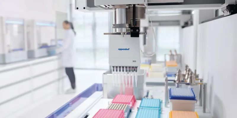
Advances in computer hardware and software, data storage and processing, optics, systems and instrumentation, labeling agents, and reagents have all contributed to the current surge in imaging in the life sciences.

Dr. Anne Carpenter leads the Imaging Platform at the Broad Institute of Harvard and MIT—a team of biologists and computer scientists who develop image analysis and data mining methods and software that are freely available to the public through the open-source CellProfiler project.
Dr. Arvind Rao has been an assistant professor in the Department of Bioinformatics and Computational Biology at the University of Texas MD Anderson Cancer Center since 2011.

Problem: In recent years, cell biology has included more emphasis on the study of rapid movement inside live cells: its dynamics, mechanisms, electrochemical signaling and protein transport. To capture these events while avoiding image artifacts, frame rates must be high enough to accurately sample these cellular phenomena. Depending on the event, these rates can range from 20 to several thousand frames per second with exposure times well below 100ms.

Imaging encompasses a wide range of techniques that enable visualization of hidden features of samples, structures, or organisms. Imaging occurs at many scales, from medical magnetic resonance imaging of patients to individual atoms. This INSIGHTS on Imaging Systems focuses on the lower end of size domains in the typical operating range of—but not limited to—microscopy.

Nongjian Tao, PhD, director of the Center for Bioelectronics and Biosensors at Arizona State University’s Biodesign Institute and professor in the School of Electrical, Computer, and Energy Engineering, talks to contributing editor Tanuja Koppal, PhD, about a new technique called plasmonic-based electrochemical microscopy (P-ECM) developed in his lab for imaging localized chemical reactions from single nanoparticles. He talks about the advantages of this technique when compared to conventional optical microscopy and scanning electrochemical microscopy (SECM) and its potential uses in diverse areas.